Structural Studies on the Empty Capsids of Physalis Mottle Virus
Krishna, S.S., Sastri, M., Savithri, H.S., Murthy, M.R.N.(2001) J Mol Biol 307: 1035
- PubMed: 11286554
- DOI: https://doi.org/10.1006/jmbi.2001.4533
- Primary Citation of Related Structures:
1E57 - PubMed Abstract:
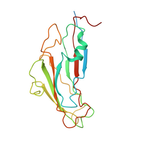
The three-dimensional crystal structure of the empty capsid of Physalis mottle tymovirus has been determined to 3.2 A resolution. The empty capsids crystallized in the space group P1, leading to 60-fold non-crystallographic redundancy. The known structure of Physalis mottle virus was used as a phasing model to initiate the structure determination by real-space electron-density averaging. The main differences between the structures of the native and the empty capsids were in residues 10 to 28 of the A-subunit, residues 1 to 9 of the B-subunit and residues 1 to 5 of the C-subunit, which are ordered only in the native virus particles. An analysis of the subunit disposition reveals that the virus has expanded radially outward by approximately 1.8 A in the empty particles. The A-subunits move in a direction that makes 10 degrees to the icosahedral 5-fold axes of symmetry. The B and C-subunits move along vectors making 12 degrees and 15 degrees to the quasi 6-fold axes. The quaternary organization of the pentameric and hexameric capsomeres are not altered significantly. However, the pentamer-hexamer contacts are reduced. Therefore, encapsidation of RNA appears to cause a reduction in the particle radius concomittant with the ordering of the N-terminal arm in the three subunits. These structural changes in Physalis mottle virus appear to be larger than the corresponding changes observed in viruses for which both the empty and full particle structures have been determined.
Organizational Affiliation:
Molecular Biophysics Unit, Indian Institute of Science, Bangalore, 560 012, India.