A refined solution structure of hen lysozyme determined using residual dipolar coupling data.
Schwalbe, H., Grimshaw, S.B., Spencer, A., Buck, M., Boyd, J., Dobson, C.M., Redfield, C., Smith, L.J.(2001) Protein Sci 10: 677-688
- PubMed: 11274458
- DOI: https://doi.org/10.1110/ps.43301
- Primary Citation of Related Structures:
1E8L - PubMed Abstract:
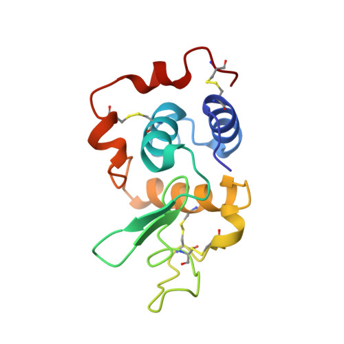
A high resolution NMR structure of hen lysozyme has been determined using 209 residual 1H-15N dipolar coupling restraints from measurements made in two different dilute liquid crystalline phases (bicelles) in conjunction with a data set of 1632 NOE distance restraints, 110 torsion angle restraints, and 60 hydrogen bond restraints. The ensemble of 50 low-energy calculated structures has an average backbone RMSD of 0.50+/-0.13A to the mean structure and of 1.49+/-0.10A to the crystal structure of hen lysozyme. To assess the importance of the dipolar coupling data in the structure determination, the final structures are compared with an ensemble calculated using an identical protocol but excluding the dipolar coupling restraints. The comparison shows that structures calculated with the dipolar coupling data are more similar to the crystal structure than those calculated without, and have better stereochemical quality. The structures also show improved quality factors when compared with additional dipolar coupling data that were not included in the structure calculations, with orientation-dependent 15N chemical shift changes measured in the bicelle solutions, and with T1/T2 values obtained from 15N relaxation measurements. Analysis of the ensemble of NMR structures and comparisons with crystal structures, 15N relaxation data, and molecular dynamics simulations of hen lysozyme provides a detailed description of the solution structure of this protein and insights into its dynamical behavior.
Organizational Affiliation:
Oxford Centre for Molecular Sciences, New Chemistry Laboratory, University of Oxford, Oxford OX1 3QT, England.