The high-resolution X-ray crystallographic structure of the ferritin (EcFtnA) of Escherichia coli; comparison with human H ferritin (HuHF) and the structures of the Fe(3+) and Zn(2+) derivatives.
Stillman, T.J., Hempstead, P.D., Artymiuk, P.J., Andrews, S.C., Hudson, A.J., Treffry, A., Guest, J.R., Harrison, P.M.(2001) J Mol Biol 307: 587-603
- PubMed: 11254384
- DOI: https://doi.org/10.1006/jmbi.2001.4475
- Primary Citation of Related Structures:
1EUM - PubMed Abstract:
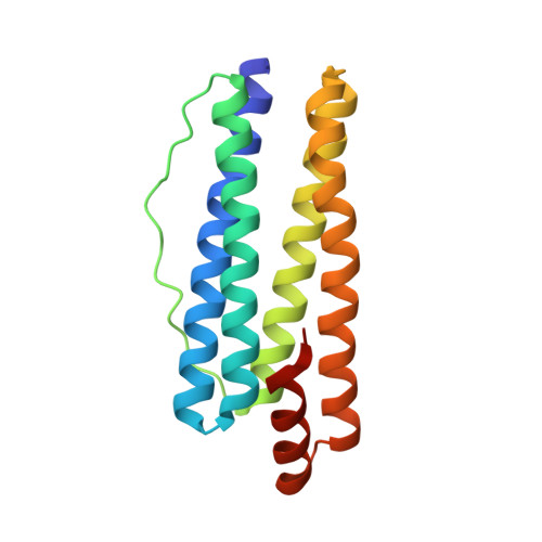
The high-resolution structure of the non-haem ferritin from Escherichia coli (EcFtnA) is presented together with those of its Fe(3+) and Zn(2+) derivatives, this being the first high-resolution X-ray analysis of the iron centres in any ferritin. The binding of both metals is accompanied by small changes in the amino acid ligand positions. Mean Fe(A)(3+)-Fe(B)(3+) and Zn(A)(2+)-Zn(B)(2+) distances are 3.24 A and 3.43 A, respectively. In both derivatives, metal ions at sites A and B are bridged by a glutamate side-chain (Glu50) in a syn-syn conformation. The Fe(3+) derivative alone shows a third metal site (Fe( C)( 3+)) joined to Fe(B)(3+) by a long anti-anti bidentate bridge through Glu130 (mean Fe(B)(3+)-Fe(C)(3+) distance 5.79 A). The third metal site is unique to the non-haem bacterial ferritins. The dinuclear site lies at the inner end of a hydrophobic channel connecting it to the outside surface of the protein shell, which may provide access for dioxygen and possibly for metal ions shielded by water. Models representing the possible binding mode of dioxygen to the dinuclear Fe(3+) pair suggest that a gauche micro-1,2 mode may be preferred stereochemically. Like those of other ferritins, the 24 subunits of EcFtnA are folded as four-helix bundles that assemble into hollow shells and both metals bind at dinuclear centres in the middle of the bundles. The structural similarity of EcFtnA to the human H chain ferritin (HuHF) is remarkable (r.m.s. deviation of main-chain atoms 0.66 A) given the low amino acid sequence identity (22 %). Many of the conserved residues are clustered at the dinuclear centre but there is very little conservation of residues making inter-subunit interactions.
Organizational Affiliation:
The Krebs Institute Department of Molecular Biology and Biotechnology, The University of Sheffield, Sheffield S10 2TN, UK.