Identification, retinoid binding, and x-ray analysis of a human retinol-binding protein.
Folli, C., Calderone, V., Ottonello, S., Bolchi, A., Zanotti, G., Stoppini, M., Berni, R.(2001) Proc Natl Acad Sci U S A 98: 3710-3715
- PubMed: 11274389
- DOI: https://doi.org/10.1073/pnas.061455898
- Primary Citation of Related Structures:
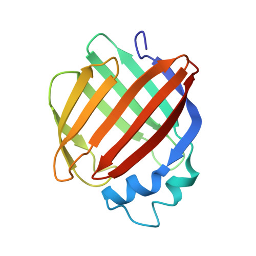
1GGL - PubMed Abstract:
Two cellular retinol-binding proteins (CRBP I and II) with distinct tissue distributions and retinoid-binding properties have been recognized thus far in mammals. Here, we report the identification of a human retinol-binding protein resembling type I (55.6% identity) and type II (49.6% identity) CRBPs, but with a unique H residue in the retinoid-binding site and a distinctively different tissue distribution. Additionally, this binding protein (CRBP III) exhibits a remarkable sequence identity (62.2%) with the recently identified iota-crystallin/CRBP of the diurnal gecko Lygodactylus picturatus [Werten, P. J. L., Röll, B., van Alten, D. M. F. & de Jong, W. W. (2000) Proc. Natl. Acad. Sci. USA 97, 3282-3287 (First Published March 21, 2000; 10.1073/pnas.050500597)]. CRBP III and all-trans-retinol form a complex (K(d) approximately 60 nM), the absorption spectrum of which is characterized by the peculiar fine structure typical of the spectra of holo-CRBP I and II. As revealed by a 2.3-A x-ray molecular model of apo-CRBP III, the amino acid residues that line the retinol-binding site in CRBP I and II are positioned nearly identically in the structure of CRBP III. At variance with the human CRBP I and II mRNAs, which are most abundant in ovary and intestine, respectively, the CRBP III mRNA is expressed at the highest levels in kidney and liver thus suggesting a prominent role for human CRBP III as an intracellular mediator of retinol metabolism in these tissues.
Organizational Affiliation:
Institute of Biochemical Sciences, University of Parma, I-43100 Parma, Italy.