1LN1

Crystal Structure of Human Phosphatidylcholine Transfer Protein in Complex with Dilinoleoylphosphatidylcholine


Experimental Data Snapshot

  • Method: X-RAY DIFFRACTION
  • Resolution: 2.40 Å
  • R-Value Free: 0.296 
  • R-Value Work: 0.234 
  • R-Value Observed: 0.237 

wwPDB Validation   3D Report Full Report


This is version 1.3 of the entry. See complete history


Literature

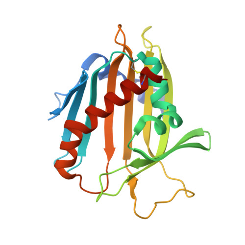
Structure of human phosphatidylcholine transfer protein in complex with its ligand.

Roderick, S.L.Chan, W.W.Agate, D.S.Olsen, L.R.Vetting, M.W.Rajashankar, K.R.Cohen, D.E.

(2002) Nat Struct Biol 9: 507-511

  • DOI: https://doi.org/10.1038/nsb812
  • Primary Citation of Related Structures:  
    1LN1, 1LN2, 1LN3

  • PubMed Abstract: 

    Phosphatidylcholines (PtdChos) comprise the most common phospholipid class in eukaryotic cells. In mammalian cells, these insoluble molecules are transferred between membranes by a highly specific phosphatidylcholine transfer protein (PC-TP) belonging to the steroidogenic acute regulatory protein related transfer (START) domain superfamily of hydrophobic ligand-binding proteins. The crystal structures of human PC-TP in complex with dilinoleoyl-PtdCho or palmitoyl-linoleoyl-PtdCho reveal that a single well-ordered PtdCho molecule occupies a centrally located tunnel. The positively charged choline headgroup of the lipid engages in cation-pi interactions within a cage formed by the faces of three aromatic residues. These binding determinants and those for the phosphoryl group may be exposed to the lipid headgroup at the membrane-water interface by a conformational change involving the amphipathic C-terminal helix and an Omega-loop. The structures presented here provide a basis for rationalizing the specificity of PC-TP for PtdCho and may identify common features used by START proteins to bind their hydrophobic ligands.


  • Organizational Affiliation

    Department of Biochemistry, Marion Bessin Liver Research Center, Albert Einstein College of Medicine, 1300 Morris Park Avenue, Bronx, New York 10461, USA. roderick@aecom.yu.edu


Macromolecules
Find similar proteins by:  (by identity cutoff)  |  3D Structure
Entity ID: 1
MoleculeChains Sequence LengthOrganismDetailsImage
Phosphatidylcholine transfer protein214Homo sapiensMutation(s): 0 
UniProt & NIH Common Fund Data Resources
Find proteins for Q9UKL6 (Homo sapiens)
Explore Q9UKL6 
Go to UniProtKB:  Q9UKL6
PHAROS:  Q9UKL6
GTEx:  ENSG00000141179 
Entity Groups  
Sequence Clusters30% Identity50% Identity70% Identity90% Identity95% Identity100% Identity
UniProt GroupQ9UKL6
Sequence Annotations
Expand
  • Reference Sequence
Small Molecules
Ligands 1 Unique
IDChains Name / Formula / InChI Key2D Diagram3D Interactions
DLP
Query on DLP

Download Ideal Coordinates CCD File 
B [auth A]1,2-DILINOLEOYL-SN-GLYCERO-3-PHOSPHOCHOLINE
C44 H80 N O8 P
FVXDQWZBHIXIEJ-LNDKUQBDSA-N
Experimental Data & Validation

Experimental Data

  • Method: X-RAY DIFFRACTION
  • Resolution: 2.40 Å
  • R-Value Free: 0.296 
  • R-Value Work: 0.234 
  • R-Value Observed: 0.237 
  • Space Group: I 4 2 2
Unit Cell:
Length ( Å )Angle ( ˚ )
a = 133.3α = 90
b = 133.3β = 90
c = 83γ = 90
Software Package:
Software NamePurpose
MAR345data collection
SCALEPACKdata scaling
X-PLORmodel building
CNSrefinement
X-PLORphasing

Structure Validation

View Full Validation Report


Entry History 

Deposition Data

Revision History  (Full details and data files)

  • Version 1.0: 2002-06-26
    Type: Initial release
  • Version 1.1: 2008-04-28
    Changes: Version format compliance
  • Version 1.2: 2011-07-13
    Changes: Version format compliance
  • Version 1.3: 2017-10-11
    Changes: Refinement description