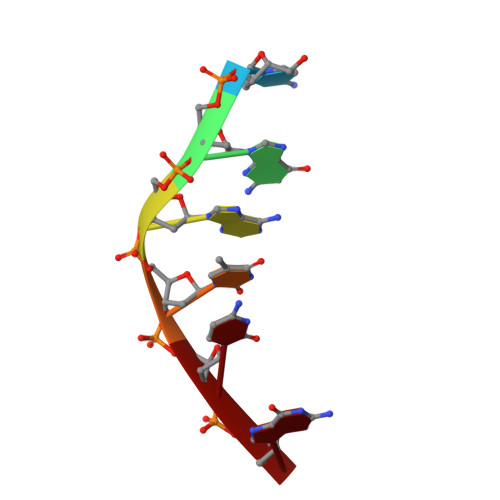
The crystal structure of the complex between a disaccharide anthracycline and the DNA hexamer d(CGATCG) reveals two different binding sites involving two DNA duplexes
Temperini, C., Messori, L., Orioli, P., Di Bugno, C., Animati, F., Ughetto, G.(2003) Nucleic Acids Res 31: 1464-1469
- PubMed: 12595554
- DOI: https://doi.org/10.1093/nar/gkg245
- Primary Citation of Related Structures:
1NAB - PubMed Abstract:
The crystal structure of the complex formed between the anthracycline antibiotic 3'-deamino-3'- hydroxy-4'-(O-L-daunosaminyl)-4-demethoxydoxo rubicin (MEN 10755), an active disaccharide analogue of doxorubicin, and the DNA hexamer d(CGATCG) has been solved to a resolution of 2.1 A. MEN 10755 exhibits a broad spectrum of antitumor activities, comparable with that of the parent compound, but there are differences in the mechanism of action as it is active in doxorubicin-resistant tumors and is more effective in stimulating topoisomerase DNA cleavage. The structure is similar to previously crystallised anthracycline- DNA complexes. However, two different binding sites arise from drug intercalation so that the two halves of the self-complementary duplex are no longer equivalent. In one site both sugar rings lie in the minor groove. In the other site the second sugar protrudes out from the DNA helix and is linked, through hydrogen bonds, to guanine of a symmetry-related DNA molecule. This is the first structure of an anthracycline-DNA complex where an interaction of the drug with a second DNA helix is observed. We discuss the present findings with respect to the relevance of the amino group for DNA binding and to the potential role played by the second sugar in the interactions with topoisomerases or other cellular targets.
Organizational Affiliation:
Department of Chemistry, University of Florence, via della Lastruccia 3, Sesto F.no (FI), Italy.