1NOA

CRYSTAL STRUCTURE OF APO-NEOCARZINOSTATIN AT 0.15 NM RESOLUTION


Experimental Data Snapshot

  • Method: X-RAY DIFFRACTION
  • Resolution: 1.50 Å
  • R-Value Observed: 0.172 

wwPDB Validation   3D Report Full Report


This is version 1.3 of the entry. See complete history


Literature

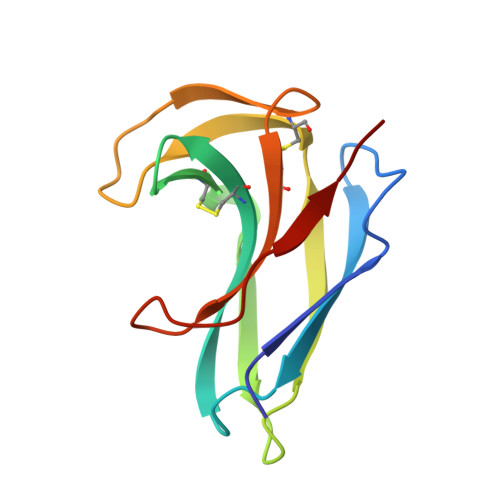
Crystal structure of apo-neocarzinostatin at 0.15-nm resolution.

Teplyakov, A.Obmolova, G.Wilson, K.Kuromizu, K.

(1993) Eur J Biochem 213: 737-741

  • DOI: https://doi.org/10.1111/j.1432-1033.1993.tb17814.x
  • Primary Citation of Related Structures:  
    1NOA

  • PubMed Abstract: 

    The three-dimensional structure of apo-neocarzinostatin, an antitumour antibiotic protein isolated from Streptomyces carzinostaticus, has been determined by X-ray diffraction at 0.15-nm resolution and refined to R = 17.2%. The crystal structure of neocarzinostatin is similar to that of the related proteins actinoxanthin and macromomycin. It is also in good agreement with the solution structure determined by NMR spectroscopy. The protein molecule consists of a seven-stranded antiparallel beta-sandwich and a smaller lobe formed by two beta-ribbons. A deep cleft between the two lobes is a putative chromophore binding site. Side chains of Trp39, Leu45, Phe52, Phe78 and the disulphide Cys37-Cys47 aligning the binding cleft in neocarzinostatin suggest the importance of hydrophobic interactions in stabilizing the chromophore molecule. Comparison of the atomic models of neocarzinostatin, actinoxanthin and macromomycin reveals functional residues which might determine specificity towards different chromophores.


  • Organizational Affiliation

    EMBL, c/o DESY, Hamburg, Germany.


Macromolecules
Find similar proteins by:  (by identity cutoff)  |  3D Structure
Entity ID: 1
MoleculeChains Sequence LengthOrganismDetailsImage
NEOCARZINOSTATIN113Streptomyces carzinostaticusMutation(s): 0 
UniProt
Find proteins for P0A3R9 (Streptomyces carzinostaticus)
Explore P0A3R9 
Go to UniProtKB:  P0A3R9
Entity Groups  
Sequence Clusters30% Identity50% Identity70% Identity90% Identity95% Identity100% Identity
UniProt GroupP0A3R9
Sequence Annotations
Expand
  • Reference Sequence
Experimental Data & Validation

Experimental Data

  • Method: X-RAY DIFFRACTION
  • Resolution: 1.50 Å
  • R-Value Observed: 0.172 
  • Space Group: P 21 21 21
Unit Cell:
Length ( Å )Angle ( ˚ )
a = 27.43α = 90
b = 34.08β = 90
c = 102.42γ = 90
Software Package:
Software NamePurpose
PROLSQrefinement

Structure Validation

View Full Validation Report


Entry History 

Deposition Data

Revision History  (Full details and data files)

  • Version 1.0: 1993-10-31
    Type: Initial release
  • Version 1.1: 2008-03-24
    Changes: Version format compliance
  • Version 1.2: 2011-07-13
    Changes: Version format compliance
  • Version 1.3: 2017-11-29
    Changes: Derived calculations, Other