1QGH

THE X-RAY STRUCTURE OF THE UNUSUAL DODECAMERIC FERRITIN FROM LISTERIA INNOCUA, REVEALS A NOVEL INTERSUBUNIT IRON BINDING SITE.


Experimental Data Snapshot

  • Method: X-RAY DIFFRACTION
  • Resolution: 2.35 Å
  • R-Value Work: 0.219 

wwPDB Validation   3D Report Full Report


This is version 1.3 of the entry. See complete history


Literature

The dodecameric ferritin from Listeria innocua contains a novel intersubunit iron-binding site.

Ilari, A.Stefanini, S.Chiancone, E.Tsernoglou, D.

(2000) Nat Struct Biol 7: 38-43

  • DOI: https://doi.org/10.1038/71236
  • Primary Citation of Related Structures:  
    1QGH

  • PubMed Abstract: 

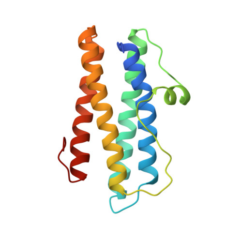
    Ferritin is characterized by a highly conserved architecture that comprises 24 subunits assembled into a spherical cage with 432 symmetry. The only known exception is the dodecameric ferritin from Listeria innocua. The structure of Listeria ferritin has been determined to a resolution of 2.35 A by molecular replacement, using as a search model the structure of Dps from Escherichia coli. The Listeria 12-mer is endowed with 23 symmetry and displays the functionally relevant structural features of the ferritin 24-mer, namely the negatively charged channels along the three-fold symmetry axes that serve for iron entry into the cavity and a negatively charged internal cavity for iron deposition. The electron density map shows 12 iron ions on the inner surface of the hollow core, at the interface between monomers related by two-fold axes. Analysis of the nature and stereochemistry of the iron-binding ligands reveals strong similarities with known ferroxidase sites. The L. innocua ferritin site, however, is the first described so far that has ligands belonging to two different subunits and is not contained within a four-helix bundle.


  • Organizational Affiliation

    CNR Center of Molecular Biology, Department of Biochemical Sciences "A. Rossi Fanelli", University La Sapienza, Piazzale Aldo Moro 5, 00185 Rome, Italy.


Macromolecules
Find similar proteins by:  (by identity cutoff)  |  3D Structure
Entity ID: 1
MoleculeChains Sequence LengthOrganismDetailsImage
NON-HEME IRON-CONTAINING FERRITIN
A, B, C, D, E
A, B, C, D, E, F, G, H, I, J, K, L
156Listeria innocuaMutation(s): 0 
UniProt
Find proteins for P80725 (Listeria innocua serovar 6a (strain ATCC BAA-680 / CLIP 11262))
Explore P80725 
Go to UniProtKB:  P80725
Entity Groups  
Sequence Clusters30% Identity50% Identity70% Identity90% Identity95% Identity100% Identity
UniProt GroupP80725
Sequence Annotations
Expand
  • Reference Sequence
Experimental Data & Validation

Experimental Data

  • Method: X-RAY DIFFRACTION
  • Resolution: 2.35 Å
  • R-Value Work: 0.219 
  • Space Group: P 21 21 21
Unit Cell:
Length ( Å )Angle ( ˚ )
a = 87.54α = 90
b = 137.5β = 90
c = 173.36γ = 90
Software Package:
Software NamePurpose
AMoREphasing
TNTrefinement
DENZOdata reduction
SCALEPACKdata scaling

Structure Validation

View Full Validation Report


Entry History 

Deposition Data

Revision History  (Full details and data files)

  • Version 1.0: 2000-01-14
    Type: Initial release
  • Version 1.1: 2008-04-26
    Changes: Version format compliance
  • Version 1.2: 2011-07-13
    Changes: Version format compliance
  • Version 1.3: 2023-08-16
    Changes: Data collection, Database references, Derived calculations, Refinement description