1RN1

THREE-DIMENSIONAL STRUCTURE OF GLN 25-RIBONUCLEASE T1 AT 1.84 ANGSTROMS RESOLUTION: STRUCTURAL VARIATIONS AT THE BASE RECOGNITION AND CATALYTIC SITES


Experimental Data Snapshot

  • Method: X-RAY DIFFRACTION
  • Resolution: 1.84 Å
  • R-Value Work: 0.144 
  • R-Value Observed: 0.144 

wwPDB Validation   3D Report Full Report


This is version 1.5 of the entry. See complete history


Literature

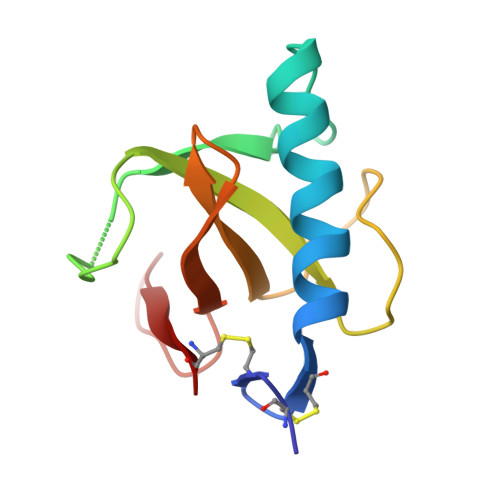
Three-dimensional structure of Gln25-ribonuclease T1 at 1.84-A resolution: structural variations at the base recognition and catalytic sites.

Arni, R.K.Pal, G.P.Ravichandran, K.G.Tulinsky, A.Walz Jr., F.G.Metcalf, P.

(1992) Biochemistry 31: 3126-3135

  • DOI: https://doi.org/10.1021/bi00127a013
  • Primary Citation of Related Structures:  
    1RN1

  • PubMed Abstract: 

    The structure of the Gln25 variant of ribonuclease T1 (RNase T1) crystallized at pH 7 and at high ionic strength has been solved by molecular replacement using the coordinates of the Lys25-RNase T1/2'-guanylic acid (2'GMP) complex at pH 5 [Arni et al. (1988) J. Biol. Chem. 263, 15358-15368] and refined by energy minimization and stereochemically restrained least-squares minimization to a crystallographic R-factor of 14.4% at 1.84-A resolution. The asymmetric unit contains three molecules, and the final model consists of 2302 protein atoms, 3 sulfates (at the catalytic sites), and 179 solvent water molecules. The estimated root mean square (rms) error in the coordinates is 0.15 A, and the rms deviation from ideality is 0.018 A for bond lengths and 1.8 degrees for bond angles. Significant differences are observed between the three molecules in the asymmetric unit at the base recognition and catalytic sites.


  • Organizational Affiliation

    European Molecular Biology Laboratory, Heidelberg, Germany.


Macromolecules
Find similar proteins by:  (by identity cutoff)  |  3D Structure
Entity ID: 1
MoleculeChains Sequence LengthOrganismDetailsImage
RIBONUCLEASE T1 ISOZYME
A, B, C
104Aspergillus oryzaeMutation(s): 0 
EC: 3.1.27.3
UniProt
Find proteins for P00651 (Aspergillus oryzae (strain ATCC 42149 / RIB 40))
Explore P00651 
Go to UniProtKB:  P00651
Entity Groups  
Sequence Clusters30% Identity50% Identity70% Identity90% Identity95% Identity100% Identity
UniProt GroupP00651
Sequence Annotations
Expand
  • Reference Sequence
Experimental Data & Validation

Experimental Data

  • Method: X-RAY DIFFRACTION
  • Resolution: 1.84 Å
  • R-Value Work: 0.144 
  • R-Value Observed: 0.144 
  • Space Group: P 21 21 21
Unit Cell:
Length ( Å )Angle ( ˚ )
a = 91.71α = 90
b = 37.54β = 90
c = 77.67γ = 90
Software Package:
Software NamePurpose
X-PLORmodel building
PROLSQrefinement
X-PLORrefinement
X-PLORphasing

Structure Validation

View Full Validation Report


Entry History 

Deposition Data

Revision History  (Full details and data files)

  • Version 1.0: 1994-01-31
    Type: Initial release
  • Version 1.1: 2008-03-03
    Changes: Version format compliance
  • Version 1.2: 2011-07-13
    Changes: Version format compliance
  • Version 1.3: 2017-11-29
    Changes: Derived calculations, Other
  • Version 1.4: 2019-07-17
    Changes: Data collection, Refinement description
  • Version 1.5: 2019-08-14
    Changes: Data collection, Refinement description