1VYO

Crystal structure of avidin


Experimental Data Snapshot

  • Method: X-RAY DIFFRACTION
  • Resolution: 1.48 Å
  • R-Value Free: 0.190 
  • R-Value Work: 0.165 
  • R-Value Observed: 0.167 

wwPDB Validation   3D Report Full Report


This is version 1.5 of the entry. See complete history


Literature

Binding Properties of Haba-Type Azo Derivatives to Avidin and Avidin-Related Protein 4.

Repo, S.Paldanius, T.A.Hytonen, V.P.Nyholm, T.K.Halling, K.K.Huuskonen, J.Pentikainen, O.T.Rissanen, K.Slotte, J.P.Airenne, T.T.Salminen, T.A.Kulomaa, M.S.Johnson, M.S.

(2006) Chem Biol 13: 1029

  • DOI: https://doi.org/10.1016/j.chembiol.2006.08.006
  • Primary Citation of Related Structures:  
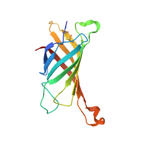
    1VYO

  • PubMed Abstract: 

    The chicken genome encodes several biotin-binding proteins, including avidin and avidin-related protein 4 (AVR4). In addition to D-biotin, avidin binds an azo dye compound, 4-hydroxyazobenzene-2-carboxylic acid (HABA), but the HABA-binding properties of AVR4 are not yet known. Differential scanning calorimetry, UV/visible spectroscopy, and molecular modeling were used to analyze the binding of 15 azo molecules to avidin and AVR4. Significant differences are seen in azo compound preferences for the two proteins, emphasizing the importance of the loop between strands beta3 and beta4 for azo ligand recognition; information on these loops is provided by the high-resolution (1.5 A) X-ray structure for avidin reported here. These results may be valuable in designing improved tools for avidin-based life science and nanobiotechnology applications.


  • Organizational Affiliation

    Department of Biochemistry and Pharmacy, Abo Akademi University, Tykistökatu 6, FI-20520 Turku, Finland.


Macromolecules
Find similar proteins by:  (by identity cutoff)  |  3D Structure
Entity ID: 1
MoleculeChains Sequence LengthOrganismDetailsImage
AVIDIN
A, B
128Gallus gallusMutation(s): 0 
UniProt
Find proteins for P02701 (Gallus gallus)
Explore P02701 
Go to UniProtKB:  P02701
Entity Groups  
Sequence Clusters30% Identity50% Identity70% Identity90% Identity95% Identity100% Identity
UniProt GroupP02701
Sequence Annotations
Expand
  • Reference Sequence
Experimental Data & Validation

Experimental Data

  • Method: X-RAY DIFFRACTION
  • Resolution: 1.48 Å
  • R-Value Free: 0.190 
  • R-Value Work: 0.165 
  • R-Value Observed: 0.167 
  • Space Group: P 21 21 2
Unit Cell:
Length ( Å )Angle ( ˚ )
a = 72.949α = 90
b = 78.772β = 90
c = 43.01γ = 90
Software Package:
Software NamePurpose
REFMACrefinement
XDSdata reduction
XDSdata scaling
AMoREphasing

Structure Validation

View Full Validation Report


Entry History 

Deposition Data

Revision History  (Full details and data files)

  • Version 1.0: 2005-07-20
    Type: Initial release
  • Version 1.1: 2011-05-08
    Changes: Version format compliance
  • Version 1.2: 2011-07-13
    Changes: Version format compliance
  • Version 1.3: 2019-07-24
    Changes: Data collection
  • Version 1.4: 2019-10-16
    Changes: Data collection, Experimental preparation
  • Version 1.5: 2023-12-13
    Changes: Data collection, Database references, Derived calculations, Refinement description