Mechanism of the Class I Kdpg Aldolase.
Fullerton, S.W.B., Griffiths, J.S., Merkel, A.B., Cheriyan, M., Wymer, N.J., Hutchins, M.J., Fierke, C.A., Toone, E.J., Naismith, J.H.(2006) Bioorg Med Chem 14: 3002
- PubMed: 16403639
- DOI: https://doi.org/10.1016/j.bmc.2005.12.022
- Primary Citation of Related Structures:
1WA3, 1WAU, 1WBH, 2C0A - PubMed Abstract:
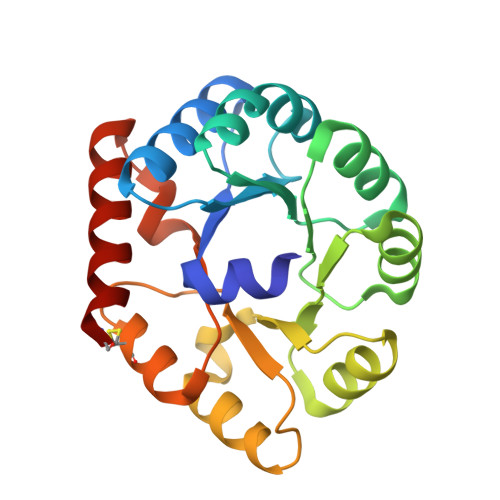
In vivo, 2-keto-3-deoxy-6-phosphogluconate (KDPG) aldolase catalyzes the reversible, stereospecific retro-aldol cleavage of KDPG to pyruvate and D-glyceraldehyde-3-phosphate. The enzyme is a lysine-dependent (Class I) aldolase that functions through the intermediacy of a Schiff base. Here, we propose a mechanism for this enzyme based on crystallographic studies of wild-type and mutant aldolases. The three dimensional structure of KDPG aldolase from the thermophile Thermotoga maritima was determined to 1.9A. The structure is the standard alpha/beta barrel observed for all Class I aldolases. At the active site Lys we observe clear density for a pyruvate Schiff base. Density for a sulfate ion bound in a conserved cluster of residues close to the Schiff base is also observed. We have also determined the structure of a mutant of Escherichia coli KDPG aldolase in which the proposed general acid/base catalyst has been removed (E45N). One subunit of the trimer contains density suggesting a trapped pyruvate carbinolamine intermediate. All three subunits contain a phosphate ion bound in a location effectively identical to that of the sulfate ion bound in the T. maritima enzyme. The sulfate and phosphate ions experimentally locate the putative phosphate binding site of the aldolase and, together with the position of the bound pyruvate, facilitate construction of a model for the full-length KDPG substrate complex. The model requires only minimal positional adjustments of the experimentally determined covalent intermediate and bound anion to accommodate full-length substrate. The model identifies the key catalytic residues of the protein and suggests important roles for two observable water molecules. The first water molecule remains bound to the enzyme during the entire catalytic cycle, shuttling protons between the catalytic glutamate and the substrate. The second water molecule arises from dehydration of the carbinolamine and serves as the nucleophilic water during hydrolysis of the enzyme-product Schiff base. The second water molecule may also mediate the base-catalyzed enolization required to form the carbon nucleophile, again bridging to the catalytic glutamate. Many aspects of this mechanism are observed in other Class I aldolases and suggest a mechanistically and, perhaps, evolutionarily related family of aldolases distinct from the N-acetylneuraminate lyase (NAL) family.
Organizational Affiliation:
Centre for Biomolecular Sciences, The University of St Andrews, St Andrews, KY16 9ST, UK.