Solution Structural Study of a DNA Duplex Containing the Guanine-N7 Adduct Formed by a Cytotoxic Platinum-Acridine Hybrid Agent
Baruah, H., Wright, M.W., Bierbach, U.(2005) Biochemistry 44: 6059-6070
- PubMed: 15835895
- DOI: https://doi.org/10.1021/bi050021b
- Primary Citation of Related Structures:
1XRW - PubMed Abstract:
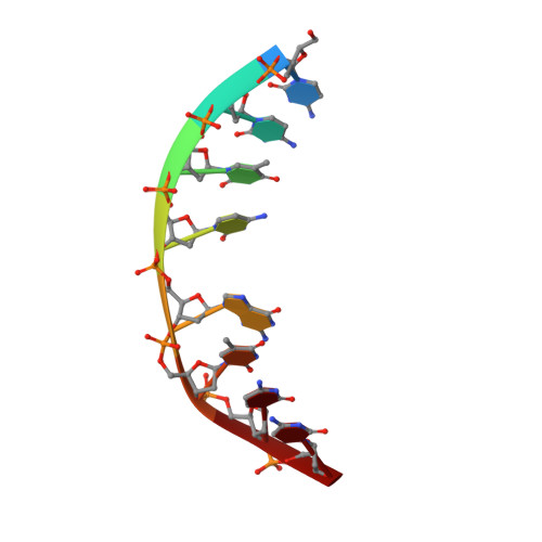
[PtCl(en)(ACRAMTU-S)](NO(3))(2) (PT-ACRAMTU; en = ethane-1,2-diamine, ACRAMTU = 1-[2-(acridin-9-ylamino)ethyl]-1,3-dimethylthiourea) is a dual metalating/intercalating DNA binding drug conjugate that shows cytotoxicity at micromolar to nanomolar concentrations in a wide range of solid tumor cell lines. In approximately 80% of its adducts, PT-ACRAMTU binds to guanine-N7 in the major groove, selectively at 5'-CG sites [Budiman, M. E. et al. (2004) Biochemistry 43, 8560-8567]. Here, we report the synthesis, physical characterization, and NMR solution structure of a site-specifically modified octamer containing this adduct, 5'-CCTCGTCC-3'/3'-GGAGCAGG-5', where the asterisk indicates the [Pt(en)ACRAMTU)](3+) fragment. The structure was determined by a combination of high-resolution 2-D NMR spectroscopy and restrained molecular dynamics/molecular mechanics (rMD/MM) calculations using 179 NOE distance restraints and refined to an r(6) weighted residual (R(x)) of 9.2 x 10(-)(2) using the complete relaxation matrix approach. An average structure was calculated from the final ensemble of 19 rMD geometries showing pairwise root-mean-square deviations of <1.05 A. The dual binding increases the thermal stability of the octamer compared to the unmodified duplex (DeltaT(m) = 13.2 degrees ). The modified sequence shows structural features reminiscent of both B- and A-type DNA. Watson-Crick hydrogen bonding is intact at and beyond the adduct site. Platinum is bound to the N7 position of G5 in the major groove, and ACRAMTU intercalates into the central 5'-C4G5/C12G13 base-pair step on the 5'-face of the platinated nucleobase. The chromophore's long axis is aligned with the long axes of the adjacent base pairs, maximizing intermolecular pi-pi stacking interactions. PT-ACRAMTU lengthens (rise, 6.62 A) and unwinds (twist, 15.4 degrees ) the duplex at the central base-pair step but does not cause helical bending. No C3'-endo deoxyribose pucker and no significant roll are observed at the site of intercalation/platination, which clearly distinguishes the PT-ACRAMTU-induced damage from the 1,2-intrastrand cross-link formed by cisplatin. Overall, the DNA perturbations produced by PT-ACRAMTU do not appear to mimic those caused by the major cisplatin lesion. Instead, intriguing structural similarities are observed for PT-ACRAMTU's monoadduct and the N7 adducts of dual major-groove alkylating/intercalating antitumor agents, such as the pluramycins.
Organizational Affiliation:
Department of Chemistry, Wake Forest University, P.O. Box 7486 Reynolda Station, Winston-Salem, North Carolina 27109, USA.