1YEW

Crystal structure of particulate methane monooxygenase


Experimental Data Snapshot

  • Method: X-RAY DIFFRACTION
  • Resolution: 2.80 Å
  • R-Value Free: 0.302 
  • R-Value Work: 0.272 
  • R-Value Observed: 0.273 

wwPDB Validation   3D Report Full Report


This is version 1.4 of the entry. See complete history


Literature

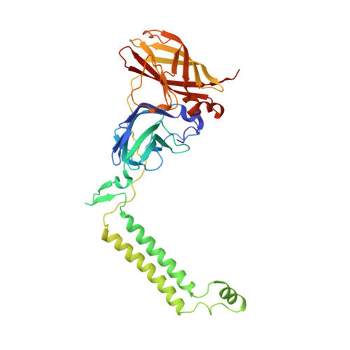
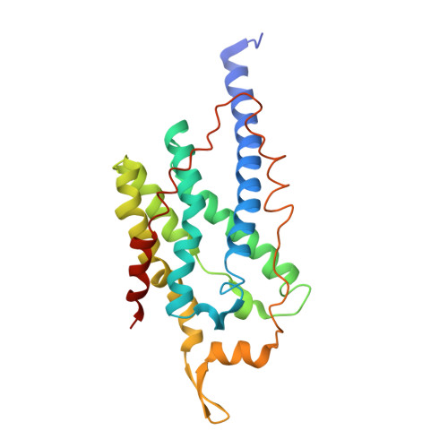
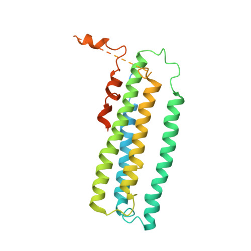
Crystal structure of a membrane-bound metalloenzyme that catalyses the biological oxidation of methane.

Lieberman, R.L.Rosenzweig, A.C.

(2005) Nature 434: 177-182

  • DOI: https://doi.org/10.1038/nature03311
  • Primary Citation of Related Structures:  
    1YEW

  • PubMed Abstract: 

    Particulate methane monooxygenase (pMMO) is an integral membrane metalloenzyme that catalyses the conversion of methane to methanol. Knowledge of how pMMO performs this extremely challenging chemistry may have an impact on the use of methane as an alternative energy source by facilitating the development of new synthetic catalysts. We have determined the structure of pMMO from the methanotroph Methylococcus capsulatus (Bath) to a resolution of 2.8 A. The enzyme is a trimer with an alpha3beta3gamma3 polypeptide arrangement. Two metal centres, modelled as mononuclear copper and dinuclear copper, are located in soluble regions of each pmoB subunit, which resembles cytochrome c oxidase subunit II. A third metal centre, occupied by zinc in the crystal, is located within the membrane. The structure provides new insight into the molecular details of biological methane oxidation.


  • Organizational Affiliation

    Department of Biochemistry, Molecular Biology, and Cell Biology, Northwestern University, Evanston, Illinois 60208, USA.


Macromolecules
Find similar proteins by:  (by identity cutoff)  |  3D Structure
Entity ID: 1
MoleculeChains Sequence LengthOrganismDetailsImage
particulate methane monooxygenase, B subunitA,
D [auth E],
G [auth I]
382Methylococcus capsulatus str. BathMutation(s): 0 
Membrane Entity: Yes 
UniProt
Find proteins for G1UBD1 (Methylococcus capsulatus (strain ATCC 33009 / NCIMB 11132 / Bath))
Explore G1UBD1 
Go to UniProtKB:  G1UBD1
Entity Groups  
Sequence Clusters30% Identity50% Identity70% Identity90% Identity95% Identity100% Identity
UniProt GroupG1UBD1
Sequence Annotations
Expand
  • Reference Sequence
Find similar proteins by:  (by identity cutoff)  |  3D Structure
Entity ID: 2
MoleculeChains Sequence LengthOrganismDetailsImage
particulate methane monooxygenase, A subunitB,
E [auth F],
H [auth J]
247Methylococcus capsulatus str. BathMutation(s): 0 
Membrane Entity: Yes 
UniProt
Find proteins for Q607G3 (Methylococcus capsulatus (strain ATCC 33009 / NCIMB 11132 / Bath))
Explore Q607G3 
Go to UniProtKB:  Q607G3
Entity Groups  
Sequence Clusters30% Identity50% Identity70% Identity90% Identity95% Identity100% Identity
UniProt GroupQ607G3
Sequence Annotations
Expand
  • Reference Sequence
Find similar proteins by:  (by identity cutoff)  |  3D Structure
Entity ID: 3
MoleculeChains Sequence LengthOrganismDetailsImage
particulate methane monooxygenase subunit C2C,
F [auth G],
I [auth K]
289Methylococcus capsulatusMutation(s): 0 
Membrane Entity: Yes 
UniProt
Find proteins for O05111 (Methylococcus capsulatus)
Explore O05111 
Go to UniProtKB:  O05111
Entity Groups  
Sequence Clusters30% Identity50% Identity70% Identity90% Identity95% Identity100% Identity
UniProt GroupO05111
Sequence Annotations
Expand
  • Reference Sequence
Small Molecules
Ligands 3 Unique
IDChains Name / Formula / InChI Key2D Diagram3D Interactions
CUA
Query on CUA

Download Ideal Coordinates CCD File 
L [auth A],
S [auth E],
V [auth I]
DINUCLEAR COPPER ION
Cu2
ALKZAGKDWUSJED-UHFFFAOYSA-N
ZN
Query on ZN

Download Ideal Coordinates CCD File 
K [auth A]
M [auth C]
N [auth C]
P [auth E]
Q [auth E]
K [auth A],
M [auth C],
N [auth C],
P [auth E],
Q [auth E],
R [auth E],
T [auth G],
W [auth K]
ZINC ION
Zn
PTFCDOFLOPIGGS-UHFFFAOYSA-N
CU
Query on CU

Download Ideal Coordinates CCD File 
J [auth A],
O [auth E],
U [auth I]
COPPER (II) ION
Cu
JPVYNHNXODAKFH-UHFFFAOYSA-N
Experimental Data & Validation

Experimental Data

  • Method: X-RAY DIFFRACTION
  • Resolution: 2.80 Å
  • R-Value Free: 0.302 
  • R-Value Work: 0.272 
  • R-Value Observed: 0.273 
  • Space Group: P 42 21 2
Unit Cell:
Length ( Å )Angle ( ˚ )
a = 264.14α = 90
b = 264.14β = 90
c = 150.005γ = 90
Software Package:
Software NamePurpose
REFMACrefinement
MAR345data collection
XDSdata scaling
SOLVEphasing

Structure Validation

View Full Validation Report


Entry History 

Deposition Data

Revision History  (Full details and data files)

  • Version 1.0: 2005-02-08
    Type: Initial release
  • Version 1.1: 2008-04-30
    Changes: Version format compliance
  • Version 1.2: 2011-07-13
    Changes: Advisory, Source and taxonomy, Version format compliance
  • Version 1.3: 2017-10-11
    Changes: Refinement description
  • Version 1.4: 2024-02-14
    Changes: Data collection, Database references, Derived calculations, Refinement description