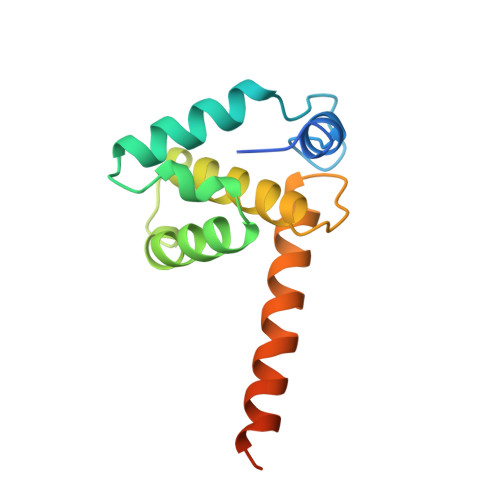
1HIW

TRIMERIC HIV-1 MATRIX PROTEIN


Experimental Data Snapshot

  • Method: X-RAY DIFFRACTION
  • Resolution: 2.30 Å
  • R-Value Free: 0.332 
  • R-Value Work: 0.259 
  • R-Value Observed: 0.259 

wwPDB Validation   3D Report Full Report


This is version 1.3 of the entry. See complete history


Literature

Crystal structures of the trimeric human immunodeficiency virus type 1 matrix protein: implications for membrane association and assembly.

Hill, C.P.Worthylake, D.Bancroft, D.P.Christensen, A.M.Sundquist, W.I.

(1996) Proc Natl Acad Sci U S A 93: 3099-3104

  • DOI: https://doi.org/10.1073/pnas.93.7.3099
  • Primary Citation of Related Structures:  
    1HIW

  • PubMed Abstract: 

    The human immunodeficiency virus type 1 (HIV-1) matrix protein forms a structural shell associated with the inner viral membrane and performs other essential functions throughout the viral life cycle. The crystal structure of the HIV-1 matrix protein, determined at 2.3 angstrom resolution, reveals that individual matrix molecules are composed of five major helices capped by a three-stranded mixed beta-sheet. Unexpectedly, the protein assembles into a trimer in three different crystal lattices, burying 1880 angstrom2 of accessible surface area at the trimer interfaces. Trimerization appears to create a large, bipartite membrane binding surface in which exposed basic residues could cooperate with the N-terminal myristoyl groups to anchor the protein on the acidic inner membrane of the virus.


  • Organizational Affiliation

    Department of Biochemistry, University of Utah, Salt Lake City, 84132, USA.


Macromolecules
Find similar proteins by:  (by identity cutoff)  |  3D Structure
Entity ID: 1
MoleculeChains Sequence LengthOrganismDetailsImage
HIV-1 MATRIX PROTEIN133Human immunodeficiency virus 1Mutation(s): 0 
UniProt
Find proteins for P12497 (Human immunodeficiency virus type 1 group M subtype B (isolate NY5))
Explore P12497 
Go to UniProtKB:  P12497
Entity Groups  
Sequence Clusters30% Identity50% Identity70% Identity90% Identity95% Identity100% Identity
UniProt GroupP12497
Sequence Annotations
Expand
  • Reference Sequence
Experimental Data & Validation

Experimental Data

  • Method: X-RAY DIFFRACTION
  • Resolution: 2.30 Å
  • R-Value Free: 0.332 
  • R-Value Work: 0.259 
  • R-Value Observed: 0.259 
  • Space Group: P 1 21 1
Unit Cell:
Length ( Å )Angle ( ˚ )
a = 64.423α = 90
b = 91.163β = 102.91
c = 73.997γ = 90
Software Package:
Software NamePurpose
X-PLORmodel building
X-PLORrefinement
DENZOdata reduction
SCALEPACKdata scaling
X-PLORphasing

Structure Validation

View Full Validation Report


Entry History 

Deposition Data

Revision History  (Full details and data files)

  • Version 1.0: 1996-10-14
    Type: Initial release
  • Version 1.1: 2008-03-24
    Changes: Version format compliance
  • Version 1.2: 2011-07-13
    Changes: Version format compliance
  • Version 1.3: 2024-02-07
    Changes: Data collection, Database references, Derived calculations, Other