1X3K

Crystal structure of a hemoglobin component (TA-V) from Tokunagayusurika akamusi


Experimental Data Snapshot

  • Method: X-RAY DIFFRACTION
  • Resolution: 1.64 Å
  • R-Value Free: 0.214 
  • R-Value Work: 0.194 

wwPDB Validation   3D Report Full Report


Ligand Structure Quality Assessment 


This is version 1.2 of the entry. See complete history


Literature

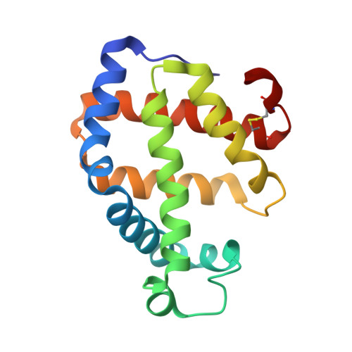
Crystal structures of two hemoglobin components from the midge larva Propsilocerus akamusi (Orthocladiinae, Diptera).

Kuwada, T.Hasegawa, T.Sato, S.Sato, I.Ishikawa, K.Takagi, T.Shishikura, F.

(2007) Gene 398: 29-34

  • DOI: https://doi.org/10.1016/j.gene.2007.02.049
  • Primary Citation of Related Structures:  
    1X3K, 1X46

  • PubMed Abstract: 

    The polymorphic components of hemoglobin (Hb) of the midge larva Propsilocerus akamusi were classified into two distinct types dependent on their spectroscopic properties, normal absorption (N) and low absorption (L). Analyses of the amino acid sequences of component VII (N-type Hb) and component V (L-type Hb) from P. akamusi indicated that one remarkable difference is the replacement of the distal histidine (His) with isoleucine (Ile) in component V. To clarify the structural differences between the two Hb components, we determined the crystal structures of components V and VII at resolutions of 1.64 A and 1.50 A, respectively. These crystal structures indicated a short additional helix comprising three amino acid residues at the C-terminal region in component V, and a typical globin fold including eight helices in component VII. Comparison of the heme regions of the Hb components suggests that the structural changes of the heme region in component V on ligation differ from that of usual Hb.


  • Organizational Affiliation

    Advanced Research Institute for the Sciences and Humanities, Nihon University, Chiyoda-ku, Tokyo 102-8251, Japan.


Macromolecules
Find similar proteins by:  (by identity cutoff)  |  3D Structure
Entity ID: 1
MoleculeChains Sequence LengthOrganismDetailsImage
hemoglobin component V152Tokunagayusurika akamusiMutation(s): 0 
UniProt
Find proteins for Q7M422 (Tokunagayusurika akamusi)
Explore Q7M422 
Go to UniProtKB:  Q7M422
Entity Groups  
Sequence Clusters30% Identity50% Identity70% Identity90% Identity95% Identity100% Identity
UniProt GroupQ7M422
Sequence Annotations
Expand
  • Reference Sequence
Small Molecules
Ligands 1 Unique
IDChains Name / Formula / InChI Key2D Diagram3D Interactions
HEM
Query on HEM

Download Ideal Coordinates CCD File 
B [auth A]PROTOPORPHYRIN IX CONTAINING FE
C34 H32 Fe N4 O4
KABFMIBPWCXCRK-RGGAHWMASA-L
Experimental Data & Validation

Experimental Data

  • Method: X-RAY DIFFRACTION
  • Resolution: 1.64 Å
  • R-Value Free: 0.214 
  • R-Value Work: 0.194 
  • Space Group: P 21 21 2
Unit Cell:
Length ( Å )Angle ( ˚ )
a = 65.144α = 90
b = 74.855β = 90
c = 33.412γ = 90
Software Package:
Software NamePurpose
CNXrefinement
CrystalCleardata reduction
CrystalCleardata scaling
CNXphasing

Structure Validation

View Full Validation Report


Ligand Structure Quality Assessment 


Entry History 

Deposition Data

Revision History  (Full details and data files)

  • Version 1.0: 2005-05-24
    Type: Initial release
  • Version 1.1: 2008-04-30
    Changes: Version format compliance
  • Version 1.2: 2011-07-13
    Changes: Version format compliance