2A3U | pdb_00002a3u

Crystal structure of sulbactam bound to E166A variant of SHV-1 beta-lactamase


Experimental Data Snapshot

  • Method: X-RAY DIFFRACTION
  • Resolution: 1.34 Å
  • R-Value Free: 
    0.194 (Depositor), 0.190 (DCC) 
  • R-Value Work: 
    0.175 (Depositor), 0.170 (DCC) 
  • R-Value Observed: 
    0.175 (Depositor) 

wwPDB Validation   3D Report Full Report


Ligand Structure Quality Assessment 

Created with Raphaël 2.3.0Worse 01 BetterLigand structure goodness of fit to experimental dataBest fitted MA4Click on this verticalbar to view detailsBest fitted TSLClick on this verticalbar to view details

This is version 1.5 of the entry. See complete history


Literature

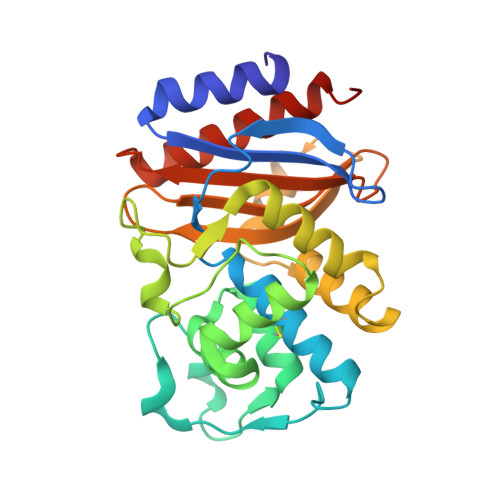
High Resolution Crystal Structures of the trans-Enamine Intermediates Formed by Sulbactam and Clavulanic Acid and E166A SHV-1 {beta}-Lactamase.

Padayatti, P.S.Helfand, M.S.Totir, M.A.Carey, M.P.Carey, P.R.Bonomo, R.A.van den Akker, F.

(2005) J Biological Chem 280: 34900-34907

  • DOI: https://doi.org/10.1074/jbc.M505333200
  • Primary Citation of Related Structures:  
    2A3U, 2A49

  • PubMed Abstract: 

    Antibiotic resistance mediated by constantly evolving beta-lactamases is a serious threat to human health. The mechanism of inhibition of these enzymes by therapeutic beta-lactamase inhibitors is probed using a novel approach involving Raman microscopy and x-ray crystallography. We have presented here the high resolution crystal structures of the beta-lactamase inhibitors sulbactam and clavulanic acid bound to the deacylation-deficient E166A variant of SHV-1 beta-lactamase. Our previous Raman measurements have identified the trans-enamine species for both inhibitors and were used to guide the soaking time and concentration to achieve full occupancy of the active sites. The two inhibitor-bound x-ray structures revealed a linear trans-enamine intermediate covalently attached to the active site Ser-70 residue. This intermediate was thought to play a key role in the transient inhibition of class A beta-lactamases. Both the Raman and x-ray data indicated that the clavulanic acid intermediate is decarboxylated. When compared with our previously determined tazobactam-bound inhibitor structure, our new inhibitor-bound structures revealed an increased disorder in the tail region of the inhibitors as well as in the enamine skeleton. The x-ray crystallographic observations correlated with the broadening of the O-C=C-N (enamine) symmetric stretch Raman band near 1595 cm(-1). Band broadening in the sulbactam and clavulanic acid inter-mediates reflected a heterogeneous conformational population that results from variations of torsional angles in the O-(C=O)-C=C=NH-C skeleton. These observations led us to conclude that the conformational stability of the trans-enamine form is critical for their transient inhibitory efficacy.


  • Organizational Affiliation

    Department of Biochemistry, Department of Chemistry, Case Western Reserve University, Cleveland, Ohio 44106, USA.


Macromolecules
Find similar proteins by:  (by identity cutoff)  |  3D Structure
Entity ID: 1
MoleculeChains Sequence LengthOrganismDetailsImage
Beta-lactamase SHV-1270Klebsiella pneumoniaeMutation(s): 1 
Gene Names: blashv1
EC: 3.5.2.6
UniProt
Find proteins for P0AD64 (Klebsiella pneumoniae)
Explore P0AD64 
Go to UniProtKB:  P0AD64
Entity Groups  
Sequence Clusters30% Identity50% Identity70% Identity90% Identity95% Identity100% Identity
UniProt GroupP0AD64
Sequence Annotations
Expand
  • Reference Sequence
Experimental Data & Validation

Experimental Data

  • Method: X-RAY DIFFRACTION
  • Resolution: 1.34 Å
  • R-Value Free:  0.194 (Depositor), 0.190 (DCC) 
  • R-Value Work:  0.175 (Depositor), 0.170 (DCC) 
  • R-Value Observed: 0.175 (Depositor) 
Space Group: P 21 21 21
Unit Cell:
Length ( Å )Angle ( ˚ )
a = 49.51α = 90
b = 55.31β = 90
c = 83.73γ = 90
Software Package:
Software NamePurpose
CNSrefinement
CCP4data scaling
CNSphasing

Structure Validation

View Full Validation Report


Ligand Structure Quality Assessment 

Created with Raphaël 2.3.0Worse 01 BetterLigand structure goodness of fit to experimental dataBest fitted MA4Click on this verticalbar to view detailsBest fitted TSLClick on this verticalbar to view details

Entry History 

Deposition Data

Revision History  (Full details and data files)

  • Version 1.0: 2005-08-02
    Type: Initial release
  • Version 1.1: 2008-04-30
    Changes: Version format compliance
  • Version 1.2: 2011-07-13
    Changes: Version format compliance
  • Version 1.3: 2017-10-11
    Changes: Refinement description
  • Version 1.4: 2021-10-20
    Changes: Database references, Derived calculations
  • Version 1.5: 2024-11-13
    Changes: Data collection, Structure summary