2BR4

cmcI-D160 Mg-SAM


Experimental Data Snapshot

  • Method: X-RAY DIFFRACTION
  • Resolution: 2.59 Å
  • R-Value Free: 0.261 
  • R-Value Work: 0.199 
  • R-Value Observed: 0.202 

wwPDB Validation   3D Report Full Report


Ligand Structure Quality Assessment 


This is version 1.2 of the entry. See complete history


Literature

Insights Into Cephamycin Biosynthesis: The Crystal Structure of Cmci from Streptomyces Clavuligerus.

Oster, L.M.Lester, D.R.Terwisscha Van Scheltinga, A.Svenda, M.Van Lun, M.Genereux, C.Andersson, I.

(2006) J Mol Biol 358: 546

  • DOI: https://doi.org/10.1016/j.jmb.2006.02.004
  • Primary Citation of Related Structures:  
    2BM8, 2BM9, 2BR3, 2BR4, 2BR5

  • PubMed Abstract: 

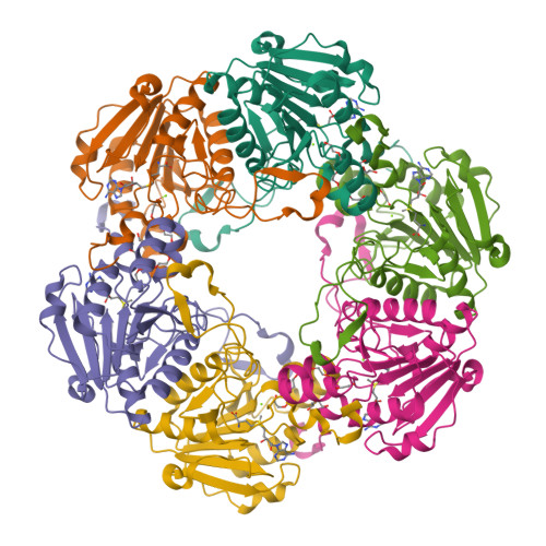
    Cephamycin C-producing microorganisms use two enzymes to convert cephalosporins to their 7alpha-methoxy derivatives. Here we report the X-ray structure of one of these enzymes, CmcI, from Streptomyces clavuligerus. The polypeptide chain of the enzyme folds into a C-terminal Rossmann domain and a smaller N-terminal domain, and the molecule packs as a hexamer in the crystal. The Rossmann domain binds S-adenosyl-L-methionine (SAM) and the demethylated product, S-adenosyl-L-homocysteine, in a fashion similar to the common binding mode of this cofactor in SAM-dependent methyltransferases. There is a magnesium-binding site in the vicinity of the SAM site with a bound magnesium ion ligated by residues Asp160, Glu186 and Asp187. The expected cephalosporin binding site near the magnesium ion is occupied by polyethyleneglycol (PEG) from the crystallisation medium. The geometry of the SAM and the magnesium binding sites is similar to that found in cathechol O-methyltransferase. The results suggest CmcI is a methyltransferase, and its most likely function is to catalyse the transfer of a methyl group from SAM to the 7alpha-hydroxy cephalosporin in the second catalytic reaction of cephamycin formation. Based on the docking of the putative substrate, 7alpha-hydroxy-O-carbamoyldeacetylcephalosporin C, to the structure of the ternary CmcI-Mg2+-SAM complex, we propose a model for substrate binding and catalysis. In this model, the 7-hydroxy group of the beta-lactam ring ligates the Mg2+ with its alpha-side facing the methyl group of SAM at a distance that would allow methylation of the hydroxyl-group.


  • Organizational Affiliation

    Department of Molecular Biology, Swedish University of Agricultural Sciences, Box 590, S-751 24 Uppsala, Sweden.


Macromolecules
Find similar proteins by:  (by identity cutoff)  |  3D Structure
Entity ID: 1
MoleculeChains Sequence LengthOrganismDetailsImage
CEPHALOSPORIN HYDROXYLASE CMCI
A, B, C, D, E
A, B, C, D, E, F
236Streptomyces clavuligerusMutation(s): 1 
UniProt
Find proteins for B5GLB3 (Streptomyces clavuligerus)
Explore B5GLB3 
Go to UniProtKB:  B5GLB3
Entity Groups  
Sequence Clusters30% Identity50% Identity70% Identity90% Identity95% Identity100% Identity
UniProt GroupB5GLB3
Sequence Annotations
Expand
  • Reference Sequence
Small Molecules
Ligands 4 Unique
IDChains Name / Formula / InChI Key2D Diagram3D Interactions
SAM
Query on SAM

Download Ideal Coordinates CCD File 
H [auth A]
L [auth B]
P [auth C]
R [auth D]
V [auth E]
H [auth A],
L [auth B],
P [auth C],
R [auth D],
V [auth E],
X [auth F]
S-ADENOSYLMETHIONINE
C15 H22 N6 O5 S
MEFKEPWMEQBLKI-FCKMPRQPSA-N
P4C
Query on P4C

Download Ideal Coordinates CCD File 
J [auth A],
N [auth B],
T [auth D]
O-ACETALDEHYDYL-HEXAETHYLENE GLYCOL
C14 H28 O8
CTLLATPOKUEFSQ-UHFFFAOYSA-N
PEG
Query on PEG

Download Ideal Coordinates CCD File 
I [auth A],
M [auth B],
S [auth D]
DI(HYDROXYETHYL)ETHER
C4 H10 O3
MTHSVFCYNBDYFN-UHFFFAOYSA-N
MG
Query on MG

Download Ideal Coordinates CCD File 
G [auth A]
K [auth B]
O [auth C]
Q [auth D]
U [auth E]
G [auth A],
K [auth B],
O [auth C],
Q [auth D],
U [auth E],
W [auth F]
MAGNESIUM ION
Mg
JLVVSXFLKOJNIY-UHFFFAOYSA-N
Experimental Data & Validation

Experimental Data

  • Method: X-RAY DIFFRACTION
  • Resolution: 2.59 Å
  • R-Value Free: 0.261 
  • R-Value Work: 0.199 
  • R-Value Observed: 0.202 
  • Space Group: P 21 21 21
Unit Cell:
Length ( Å )Angle ( ˚ )
a = 91.541α = 90
b = 102.364β = 90
c = 181.905γ = 90
Software Package:
Software NamePurpose
REFMACrefinement
DENZOdata reduction
SCALEPACKdata scaling
MOLREPphasing

Structure Validation

View Full Validation Report


Ligand Structure Quality Assessment 


Entry History 

Deposition Data

Revision History  (Full details and data files)

  • Version 1.0: 2006-03-15
    Type: Initial release
  • Version 1.1: 2011-07-13
    Changes: Advisory, Version format compliance
  • Version 1.2: 2023-12-13
    Changes: Data collection, Database references, Derived calculations, Other, Refinement description