2CJP

Structure of potato (Solanum tuberosum) epoxide hydrolase I (StEH1)


Experimental Data Snapshot

  • Method: X-RAY DIFFRACTION
  • Resolution: 1.95 Å
  • R-Value Free: 0.240 
  • R-Value Work: 0.203 
  • R-Value Observed: 0.205 

wwPDB Validation   3D Report Full Report


This is version 1.4 of the entry. See complete history


Literature

X-Ray Structure of Potato Epoxide Hydrolase Sheds Light on Substrate Specificity in Plant Enzymes.

Mowbray, S.L.Elfstrom, L.T.Ahlgren, K.M.Andersson, C.E.Widersten, M.

(2006) Protein Sci 15: 1628

  • DOI: https://doi.org/10.1110/ps.051792106
  • Primary Citation of Related Structures:  
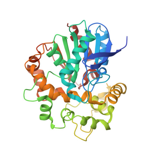
    2CJP

  • PubMed Abstract: 

    Epoxide hydrolases catalyze the conversion of epoxides to diols. The known functions of such enzymes include detoxification of xenobiotics, drug metabolism, synthesis of signaling compounds, and intermediary metabolism. In plants, epoxide hydrolases are thought to participate in general defense systems. In the present study, we report the first structure of a plant epoxide hydrolase, one of the four homologous enzymes found in potato. The structure was solved by molecular replacement and refined to a resolution of 1.95 A. Analysis of the structure allows a better understanding of the observed substrate specificities and activity. Further, comparisons with mammalian and fungal epoxide hydrolase structures reported earlier show the basis of differing substrate specificities in the various epoxide hydrolase subfamilies. Most plant enzymes, like the potato epoxide hydrolase, are expected to be monomers with a preference for substrates with long lipid-like substituents of the epoxide ring. The significance of these results in the context of biological roles and industrial applications is discussed.


  • Organizational Affiliation

    Department of Molecular Biology, Swedish University of Agricultural Sciences, Uppsala, Sweden. mowbray@xray.bmc.uu.se


Macromolecules
Find similar proteins by:  (by identity cutoff)  |  3D Structure
Entity ID: 1
MoleculeChains Sequence LengthOrganismDetailsImage
EPOXIDE HYDROLASE
A, B
328Solanum tuberosumMutation(s): 0 
EC: 3.3.2.3
UniProt
Find proteins for Q41415 (Solanum tuberosum)
Explore Q41415 
Go to UniProtKB:  Q41415
Entity Groups  
Sequence Clusters30% Identity50% Identity70% Identity90% Identity95% Identity100% Identity
UniProt GroupQ41415
Sequence Annotations
Expand
  • Reference Sequence
Small Molecules
Binding Affinity Annotations 
IDSourceBinding Affinity
VPR Binding MOAD:  2CJP Ki: 8.00e+5 (nM) from 1 assay(s)
Experimental Data & Validation

Experimental Data

  • Method: X-RAY DIFFRACTION
  • Resolution: 1.95 Å
  • R-Value Free: 0.240 
  • R-Value Work: 0.203 
  • R-Value Observed: 0.205 
  • Space Group: P 21 21 21
Unit Cell:
Length ( Å )Angle ( ˚ )
a = 55.926α = 90
b = 98.532β = 90
c = 122.033γ = 90
Software Package:
Software NamePurpose
REFMACrefinement
MOSFLMdata reduction
SCALAdata scaling
MOLREPphasing

Structure Validation

View Full Validation Report


Entry History 

Deposition Data

Revision History  (Full details and data files)

  • Version 1.0: 2006-06-07
    Type: Initial release
  • Version 1.1: 2011-05-08
    Changes: Version format compliance
  • Version 1.2: 2011-07-13
    Changes: Version format compliance
  • Version 1.3: 2019-05-15
    Changes: Data collection, Experimental preparation, Other
  • Version 1.4: 2023-12-13
    Changes: Data collection, Database references, Other, Refinement description