2F4K

Chicken villin subdomain HP-35, K65(NLE), N68H, K70(NLE), PH9


Experimental Data Snapshot

  • Method: X-RAY DIFFRACTION
  • Resolution: 1.05 Å
  • R-Value Free: 0.166 
  • R-Value Observed: 0.140 

wwPDB Validation   3D Report Full Report


This is version 1.5 of the entry. See complete history


Literature

Sub-microsecond Protein Folding.

Kubelka, J.Chiu, T.K.Davies, D.R.Eaton, W.A.Hofrichter, J.

(2006) J Mol Biol 359: 546-553

  • DOI: https://doi.org/10.1016/j.jmb.2006.03.034
  • Primary Citation of Related Structures:  
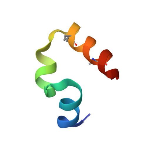
    2F4K

  • PubMed Abstract: 

    We have investigated the structure, equilibria, and folding kinetics of an engineered 35-residue subdomain of the chicken villin headpiece, an ultrafast-folding protein. Substitution of two buried lysine residues by norleucine residues stabilizes the protein by 1 kcal/mol and increases the folding rate sixfold, as measured by nanosecond laser T-jump. The folding rate at 300 K is (0.7 micros)(-1) with little or no temperature dependence, making this protein the first sub-microsecond folder, with a rate only twofold slower than the theoretically predicted speed limit. Using the 70 ns process to obtain the effective diffusion coefficient, the free energy barrier height is estimated from Kramers theory to be less than approximately 1 kcal/mol. X-ray crystallographic determination at 1A resolution shows no significant change in structure compared to the single-norleucine-substituted molecule and suggests that the increased stability is electrostatic in origin. The ultrafast folding rate, very accurate X-ray structure, and small size make this engineered villin subdomain an ideal system for simulation by atomistic molecular dynamics with explicit solvent.


  • Organizational Affiliation

    Laboratory of Chemical Physics, National Institute of Diabetes and Digestive and Kidney Diseases, National Institutes of Health, Bethesda, MD 20892-0520, USA.


Macromolecules
Find similar proteins by:  (by identity cutoff)  |  3D Structure
Entity ID: 1
MoleculeChains Sequence LengthOrganismDetailsImage
Villin-135N/AMutation(s): 3 
UniProt
Find proteins for P02640 (Gallus gallus)
Explore P02640 
Go to UniProtKB:  P02640
Entity Groups  
Sequence Clusters30% Identity50% Identity70% Identity90% Identity95% Identity100% Identity
UniProt GroupP02640
Sequence Annotations
Expand
  • Reference Sequence
Small Molecules
Modified Residues  1 Unique
IDChains TypeFormula2D DiagramParent
NLE
Query on NLE
A
L-PEPTIDE LINKINGC6 H13 N O2LEU
Experimental Data & Validation

Experimental Data

  • Method: X-RAY DIFFRACTION
  • Resolution: 1.05 Å
  • R-Value Free: 0.166 
  • R-Value Observed: 0.140 
  • Space Group: C 2 2 21
Unit Cell:
Length ( Å )Angle ( ˚ )
a = 19.677α = 90
b = 39.901β = 90
c = 75.089γ = 90
Software Package:
Software NamePurpose
HKL-2000data collection
HKL-2000data reduction
SHELXmodel building
SHELXL-97refinement
HKL-2000data reduction
HKL-2000data scaling
SHELXphasing

Structure Validation

View Full Validation Report


Entry History 

Deposition Data

Revision History  (Full details and data files)

  • Version 1.0: 2006-04-11
    Type: Initial release
  • Version 1.1: 2008-05-01
    Changes: Version format compliance
  • Version 1.2: 2011-07-13
    Changes: Version format compliance
  • Version 1.3: 2018-03-14
    Changes: Database references
  • Version 1.4: 2023-08-23
    Changes: Data collection, Database references, Derived calculations, Refinement description
  • Version 1.5: 2023-11-15
    Changes: Data collection