2JDI

Ground state structure of F1-ATPase from bovine heart mitochondria (Bovine F1-ATPase crystallised in the absence of azide)


Experimental Data Snapshot

  • Method: X-RAY DIFFRACTION
  • Resolution: 1.90 Å
  • R-Value Free: 0.220 
  • R-Value Work: 0.175 
  • R-Value Observed: 0.177 

wwPDB Validation   3D Report Full Report


Ligand Structure Quality Assessment 


This is version 1.2 of the entry. See complete history


Literature

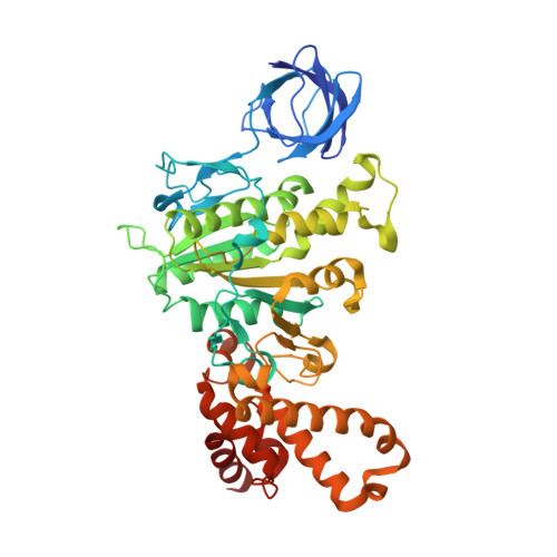
Ground State Structure of F1-ATPase from Bovine Heart Mitochondria at 1.9 A Resolution

Bowler, M.W.Montgomery, M.G.Leslie, A.G.W.Walker, J.E.

(2007) J Biol Chem 282: 14238

  • DOI: https://doi.org/10.1074/jbc.M700203200
  • Primary Citation of Related Structures:  
    2JDI

  • PubMed Abstract: 

    The structure of bovine F(1)-ATPase, crystallized in the presence of AMP-PNP and ADP, but in the absence of azide, has been determined at 1.9A resolution. This structure has been compared with the previously described structure of bovine F(1)-ATPase determined at 1.95A resolution with crystals grown under the same conditions but in the presence of azide. The two structures are extremely similar, but they differ in the nucleotides that are bound to the catalytic site in the beta(DP)-subunit. In the present structure, the nucleotide binding sites in the beta(DP)- and beta(TP)-subunits are both occupied by AMP-PNP, whereas in the earlier structure, the beta(TP) site was occupied by AMP-PNP and the beta(DP) site by ADP, where its binding is enhanced by a bound azide ion. Also, the conformation of the side chain of the catalytically important residue, alphaArg-373 differs in the beta(DP)- and beta(TP)-subunits. Thus, the structure with bound azide represents the ADP inhibited state of the enzyme, and the new structure represents a ground state intermediate in the active catalytic cycle of ATP hydrolysis.


  • Organizational Affiliation

    The Medical Research Council Dunn Human Nutrition Unit, Hills Road, Cambridge, UK.


Macromolecules
Find similar proteins by:  (by identity cutoff)  |  3D Structure
Entity ID: 1
MoleculeChains Sequence LengthOrganismDetailsImage
ATP SYNTHASE SUBUNIT ALPHA HEART ISOFORM
A, B, C
510Bos taurusMutation(s): 0 
EC: 3.6.3.14
Membrane Entity: Yes 
UniProt
Find proteins for P19483 (Bos taurus)
Explore P19483 
Go to UniProtKB:  P19483
Entity Groups  
Sequence Clusters30% Identity50% Identity70% Identity90% Identity95% Identity100% Identity
UniProt GroupP19483
Sequence Annotations
Expand
  • Reference Sequence
Find similar proteins by:  (by identity cutoff)  |  3D Structure
Entity ID: 2
MoleculeChains Sequence LengthOrganismDetailsImage
ATP SYNTHASE SUBUNIT BETA
D, E, F
482Bos taurusMutation(s): 0 
EC: 3.6.3.14
Membrane Entity: Yes 
UniProt
Find proteins for P00829 (Bos taurus)
Explore P00829 
Go to UniProtKB:  P00829
Entity Groups  
Sequence Clusters30% Identity50% Identity70% Identity90% Identity95% Identity100% Identity
UniProt GroupP00829
Sequence Annotations
Expand
  • Reference Sequence
Find similar proteins by:  (by identity cutoff)  |  3D Structure
Entity ID: 3
MoleculeChains Sequence LengthOrganismDetailsImage
ATP SYNTHASE GAMMA CHAIN273Bos taurusMutation(s): 0 
Membrane Entity: Yes 
UniProt
Find proteins for P05631 (Bos taurus)
Explore P05631 
Go to UniProtKB:  P05631
Entity Groups  
Sequence Clusters30% Identity50% Identity70% Identity90% Identity95% Identity100% Identity
UniProt GroupP05631
Sequence Annotations
Expand
  • Reference Sequence
Find similar proteins by:  (by identity cutoff)  |  3D Structure
Entity ID: 4
MoleculeChains Sequence LengthOrganismDetailsImage
ATP SYNTHASE DELTA CHAIN146Bos taurusMutation(s): 0 
Membrane Entity: Yes 
UniProt
Find proteins for P05630 (Bos taurus)
Explore P05630 
Go to UniProtKB:  P05630
Entity Groups  
Sequence Clusters30% Identity50% Identity70% Identity90% Identity95% Identity100% Identity
UniProt GroupP05630
Sequence Annotations
Expand
  • Reference Sequence
Find similar proteins by:  (by identity cutoff)  |  3D Structure
Entity ID: 5
MoleculeChains Sequence LengthOrganismDetailsImage
ATP SYNTHASE EPSILON CHAIN50Bos taurusMutation(s): 0 
Membrane Entity: Yes 
UniProt
Find proteins for P05632 (Bos taurus)
Explore P05632 
Go to UniProtKB:  P05632
Entity Groups  
Sequence Clusters30% Identity50% Identity70% Identity90% Identity95% Identity100% Identity
UniProt GroupP05632
Sequence Annotations
Expand
  • Reference Sequence
Small Molecules
Ligands 2 Unique
IDChains Name / Formula / InChI Key2D Diagram3D Interactions
ANP
Query on ANP

Download Ideal Coordinates CCD File 
J [auth A],
L [auth B],
N [auth C],
P [auth D],
R [auth F]
PHOSPHOAMINOPHOSPHONIC ACID-ADENYLATE ESTER
C10 H17 N6 O12 P3
PVKSNHVPLWYQGJ-KQYNXXCUSA-N
MG
Query on MG

Download Ideal Coordinates CCD File 
K [auth A],
M [auth B],
O [auth C],
Q [auth D],
S [auth F]
MAGNESIUM ION
Mg
JLVVSXFLKOJNIY-UHFFFAOYSA-N
Experimental Data & Validation

Experimental Data

  • Method: X-RAY DIFFRACTION
  • Resolution: 1.90 Å
  • R-Value Free: 0.220 
  • R-Value Work: 0.175 
  • R-Value Observed: 0.177 
  • Space Group: P 21 21 21
Unit Cell:
Length ( Å )Angle ( ˚ )
a = 105.61α = 90
b = 123.13β = 90
c = 261.76γ = 90
Software Package:
Software NamePurpose
REFMACrefinement
MOSFLMdata reduction
SCALAdata scaling
MOLREPphasing

Structure Validation

View Full Validation Report


Ligand Structure Quality Assessment 


Entry History 

Deposition Data

Revision History  (Full details and data files)

  • Version 1.0: 2007-03-13
    Type: Initial release
  • Version 1.1: 2011-07-13
    Changes: Refinement description, Version format compliance
  • Version 1.2: 2023-12-13
    Changes: Data collection, Database references, Derived calculations, Other, Refinement description