2ONK

ABC transporter ModBC in complex with its binding protein ModA


Experimental Data Snapshot

  • Method: X-RAY DIFFRACTION
  • Resolution: 3.10 Å
  • R-Value Free: 0.283 
  • R-Value Work: 0.256 
  • R-Value Observed: 0.256 

wwPDB Validation   3D Report Full Report


This is version 1.3 of the entry. See complete history


Literature

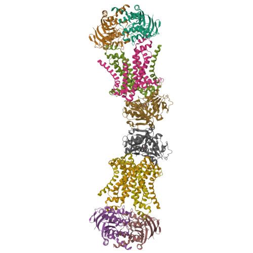
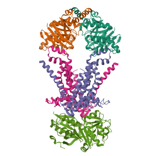
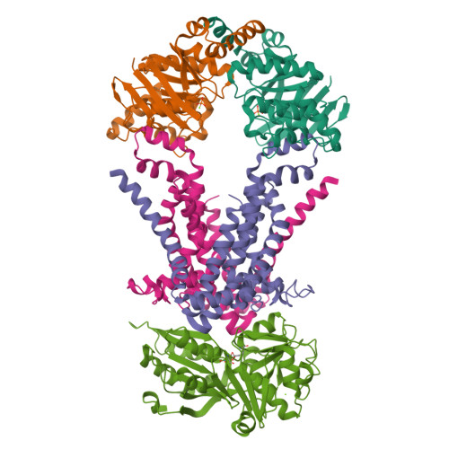
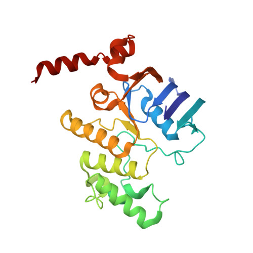
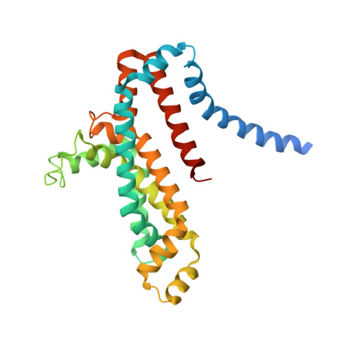
Structure of an ABC transporter in complex with its binding protein.

Hollenstein, K.Frei, D.C.Locher, K.P.

(2007) Nature 446: 213-216

  • DOI: https://doi.org/10.1038/nature05626
  • Primary Citation of Related Structures:  
    2ONK, 2ONR, 2ONS

  • PubMed Abstract: 

    ATP-binding cassette (ABC) transporter proteins carry diverse substrates across cell membranes. Whereas clinically relevant ABC exporters are implicated in various diseases or cause multidrug resistance of cancer cells, bacterial ABC importers are essential for the uptake of nutrients, including rare elements such as molybdenum. A detailed understanding of their mechanisms requires direct visualization at high resolution and in distinct conformations. Our recent structure of the multidrug ABC exporter Sav1866 has revealed an outward-facing conformation of the transmembrane domains coupled to a closed conformation of the nucleotide-binding domains, reflecting the ATP-bound state. Here we present the 3.1 A crystal structure of a putative molybdate transporter (ModB2C2) from Archaeoglobus fulgidus in complex with its binding protein (ModA). Twelve transmembrane helices of the ModB subunits provide an inward-facing conformation, with a closed gate near the external membrane boundary. The ATP-hydrolysing ModC subunits reveal a nucleotide-free, open conformation, whereas the attached binding protein aligns the substrate-binding cleft with the entrance to the presumed translocation pathway. Structural comparison of ModB2C2A with Sav1866 suggests a common alternating access and release mechanism, with binding of ATP promoting an outward-facing conformation and dissociation of the hydrolysis products promoting an inward-facing conformation.


  • Organizational Affiliation

    Institute of Molecular Biology and Biophysics, ETH Zurich, 8093 Zurich, Switzerland.


Macromolecules
Find similar proteins by:  (by identity cutoff)  |  3D Structure
Entity ID: 1
MoleculeChains Sequence LengthOrganismDetailsImage
Molybdate/tungstate ABC transporter, ATP-binding proteinA,
B,
C [auth F],
D [auth G]
240Archaeoglobus fulgidusMutation(s): 0 
Membrane Entity: Yes 
UniProt
Find proteins for O30144 (Archaeoglobus fulgidus (strain ATCC 49558 / DSM 4304 / JCM 9628 / NBRC 100126 / VC-16))
Explore O30144 
Go to UniProtKB:  O30144
Entity Groups  
Sequence Clusters30% Identity50% Identity70% Identity90% Identity95% Identity100% Identity
UniProt GroupO30144
Sequence Annotations
Expand
  • Reference Sequence
Find similar proteins by:  (by identity cutoff)  |  3D Structure
Entity ID: 2
MoleculeChains Sequence LengthOrganismDetailsImage
Molybdate/tungstate ABC transporter, permease proteinE [auth C],
F [auth D],
G [auth H],
H [auth I]
284Archaeoglobus fulgidusMutation(s): 0 
Membrane Entity: Yes 
UniProt
Find proteins for O30143 (Archaeoglobus fulgidus (strain ATCC 49558 / DSM 4304 / JCM 9628 / NBRC 100126 / VC-16))
Explore O30143 
Go to UniProtKB:  O30143
Entity Groups  
Sequence Clusters30% Identity50% Identity70% Identity90% Identity95% Identity100% Identity
UniProt GroupO30143
Sequence Annotations
Expand
  • Reference Sequence
Find similar proteins by:  (by identity cutoff)  |  3D Structure
Entity ID: 3
MoleculeChains Sequence LengthOrganismDetailsImage
Molybdate/tungstate binding proteinI [auth E],
J
314Archaeoglobus fulgidusMutation(s): 0 
Membrane Entity: Yes 
UniProt
Find proteins for O30142 (Archaeoglobus fulgidus (strain ATCC 49558 / DSM 4304 / JCM 9628 / NBRC 100126 / VC-16))
Explore O30142 
Go to UniProtKB:  O30142
Entity Groups  
Sequence Clusters30% Identity50% Identity70% Identity90% Identity95% Identity100% Identity
UniProt GroupO30142
Sequence Annotations
Expand
  • Reference Sequence
Small Molecules
Ligands 3 Unique
IDChains Name / Formula / InChI Key2D Diagram3D Interactions
WO4
Query on WO4

Download Ideal Coordinates CCD File 
S [auth E],
U [auth J]
TUNGSTATE(VI)ION
O4 W
PBYZMCDFOULPGH-UHFFFAOYSA-N
PO4
Query on PO4

Download Ideal Coordinates CCD File 
K [auth A],
M [auth B],
O [auth F],
Q [auth G]
PHOSPHATE ION
O4 P
NBIIXXVUZAFLBC-UHFFFAOYSA-K
MG
Query on MG

Download Ideal Coordinates CCD File 
L [auth A]
N [auth B]
P [auth F]
R [auth G]
T [auth E]
L [auth A],
N [auth B],
P [auth F],
R [auth G],
T [auth E],
V [auth J]
MAGNESIUM ION
Mg
JLVVSXFLKOJNIY-UHFFFAOYSA-N
Experimental Data & Validation

Experimental Data

  • Method: X-RAY DIFFRACTION
  • Resolution: 3.10 Å
  • R-Value Free: 0.283 
  • R-Value Work: 0.256 
  • R-Value Observed: 0.256 
  • Space Group: P 1 21 1
Unit Cell:
Length ( Å )Angle ( ˚ )
a = 93.032α = 90
b = 171.203β = 98.79
c = 158.47γ = 90
Software Package:
Software NamePurpose
CNSrefinement
PDB_EXTRACTdata extraction

Structure Validation

View Full Validation Report


Entry History 

Deposition Data

Revision History  (Full details and data files)

  • Version 1.0: 2007-03-06
    Type: Initial release
  • Version 1.1: 2008-05-01
    Changes: Version format compliance
  • Version 1.2: 2011-07-13
    Changes: Version format compliance
  • Version 1.3: 2023-12-27
    Changes: Data collection, Database references, Derived calculations