2VXN

E65Q-TIM complexed with phosphoglycolohydroxamate at 0.82 A resolution


Experimental Data Snapshot

  • Method: X-RAY DIFFRACTION
  • Resolution: 0.82 Å
  • R-Value Free: 0.103 
  • R-Value Observed: 0.092 

wwPDB Validation   3D Report Full Report


Ligand Structure Quality Assessment 


This is version 1.4 of the entry. See complete history


Literature

Atomic Resolution Crystallography of a Complex of Triosephosphate Isomerase with a Reaction-Intermediate Analog: New Insight in the Proton Transfer Reaction Mechanism.

Alahuhta, M.Wierenga, R.K.

(2010) Proteins 78: 1878

  • DOI: https://doi.org/10.1002/prot.22701
  • Primary Citation of Related Structures:  
    2VXN

  • PubMed Abstract: 

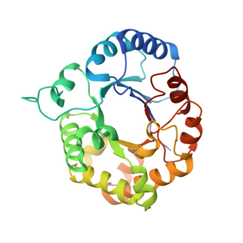
    Enzymes achieve their catalytic proficiency by precisely positioning the substrate and catalytic residues with respect to each other. Atomic resolution crystallography is an excellent tool to study the important details of these geometric active-site features. Here, we have investigated the reaction mechanism of triosephosphate isomerase (TIM) using atomic resolution crystallographic studies at 0.82-A resolution of leishmanial TIM complexed with the well-studied reaction-intermediate analog phosphoglycolohydroxamate (PGH). Remaining unresolved aspects of the reaction mechanism of TIM such as the protonation state of the first reaction intermediate and the properties of the hydrogen-bonding interactions in the active site are being addressed. The hydroxamate moiety of PGH interacts via unusually short hydrogen bonds of its N1-O1 moiety with the carboxylate group of the catalytic glutamate (Glu167), for example, the distance of N1(PGH)-OE2(Glu167) is 2.69 +/- 0.01 A and the distance of O1(PGH)-OE1(Glu167) is 2.60 +/- 0.01 A. Structural comparisons show that the side chain of the catalytic base (Glu167) can move during the reaction cycle in a small cavity, located above the hydroxamate plane. The structure analysis suggests that the hydroxamate moiety of PGH is negatively charged. Therefore, the bound PGH mimics the negatively charged enediolate intermediate, which is formed immediately after the initial proton abstraction from DHAP by the catalytic glutamate. The new findings are discussed in the context of the current knowledge of the TIM reaction mechanism.


  • Organizational Affiliation

    Department of Biochemistry, Biocenter Oulu, University of Oulu, Oulu, Finland.


Macromolecules
Find similar proteins by:  (by identity cutoff)  |  3D Structure
Entity ID: 1
MoleculeChains Sequence LengthOrganismDetailsImage
TRIOSEPHOSPHATE ISOMERASE251Leishmania mexicanaMutation(s): 1 
EC: 5.3.1.1
UniProt
Find proteins for P48499 (Leishmania mexicana)
Explore P48499 
Go to UniProtKB:  P48499
Entity Groups  
Sequence Clusters30% Identity50% Identity70% Identity90% Identity95% Identity100% Identity
UniProt GroupP48499
Sequence Annotations
Expand
  • Reference Sequence
Small Molecules
Ligands 4 Unique
IDChains Name / Formula / InChI Key2D Diagram3D Interactions
PGH
Query on PGH

Download Ideal Coordinates CCD File 
B [auth A]PHOSPHOGLYCOLOHYDROXAMIC ACID
C2 H6 N O6 P
BAXHHWZKQZIJID-UHFFFAOYSA-N
PGA
Query on PGA

Download Ideal Coordinates CCD File 
C [auth A]2-PHOSPHOGLYCOLIC ACID
C2 H5 O6 P
ASCFNMCAHFUBCO-UHFFFAOYSA-N
GOL
Query on GOL

Download Ideal Coordinates CCD File 
D [auth A],
E [auth A],
F [auth A],
G [auth A],
H [auth A]
GLYCEROL
C3 H8 O3
PEDCQBHIVMGVHV-UHFFFAOYSA-N
ACT
Query on ACT

Download Ideal Coordinates CCD File 
I [auth A]ACETATE ION
C2 H3 O2
QTBSBXVTEAMEQO-UHFFFAOYSA-M
Binding Affinity Annotations 
IDSourceBinding Affinity
PGH PDBBind:  2VXN Kd: 1.00e+4 (nM) from 1 assay(s)
Experimental Data & Validation

Experimental Data

  • Method: X-RAY DIFFRACTION
  • Resolution: 0.82 Å
  • R-Value Free: 0.103 
  • R-Value Observed: 0.092 
  • Space Group: C 1 2 1
Unit Cell:
Length ( Å )Angle ( ˚ )
a = 98.36α = 90
b = 50.65β = 117.56
c = 58.66γ = 90
Software Package:
Software NamePurpose
SHELXL-97refinement
XDSdata reduction
XDSdata scaling
MOLREPphasing

Structure Validation

View Full Validation Report


Ligand Structure Quality Assessment 


Entry History 

Deposition Data

Revision History  (Full details and data files)

  • Version 1.0: 2009-07-14
    Type: Initial release
  • Version 1.1: 2011-05-07
    Changes: Version format compliance
  • Version 1.2: 2011-07-13
    Changes: Version format compliance
  • Version 1.3: 2019-07-24
    Changes: Data collection
  • Version 1.4: 2023-12-13
    Changes: Data collection, Database references, Derived calculations, Other, Refinement description