2WHE

Structure of native Beta-Phosphoglucomutase in an open conformation without bound ligands.


Experimental Data Snapshot

  • Method: X-RAY DIFFRACTION
  • Resolution: 1.55 Å
  • R-Value Free: 0.208 
  • R-Value Work: 0.176 
  • R-Value Observed: 0.177 

wwPDB Validation   3D Report Full Report


This is version 1.3 of the entry. See complete history


Literature

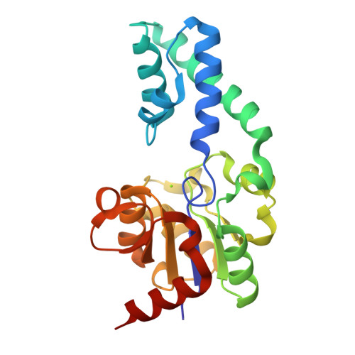
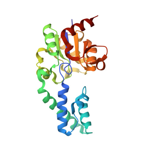
Atomic Details of Near-Transition State Conformers for Enzyme Phosphoryl Transfer Revealed by Mgf-3 Rather Than by Phosphoranes.

Baxter, N.J.Bowler, M.W.Alizadeh, T.Cliff, M.J.Hounslow, A.M.Wu, B.Berkowitz, D.B.Williams, N.H.Blackburn, G.M.Waltho, J.P.

(2010) Proc Natl Acad Sci U S A 107: 4555

  • DOI: https://doi.org/10.1073/pnas.0910333106
  • Primary Citation of Related Structures:  
    2WF5, 2WF6, 2WHE

  • PubMed Abstract: 

    Prior evidence supporting the direct observation of phosphorane intermediates in enzymatic phosphoryl transfer reactions was based on the interpretation of electron density corresponding to trigonal species bridging the donor and acceptor atoms. Close examination of the crystalline state of beta-phosphoglucomutase, the archetypal phosphorane intermediate-containing enzyme, reveals that the trigonal species is not PO-3 , but is MgF-3 (trifluoromagnesate). Although MgF-3 complexes are transition state analogues rather than phosphoryl group transfer reaction intermediates, the presence of fluorine nuclei in near-transition state conformations offers new opportunities to explore the nature of the interactions, in particular the independent measures of local electrostatic and hydrogen-bonding distributions using 19F NMR. Measurements on three beta-PGM-MgF-3 -sugar phosphate complexes show a remarkable relationship between NMR chemical shifts, primary isotope shifts, NOEs, cross hydrogen bond F...H-N scalar couplings, and the atomic positions determined from the high-resolution crystal structure of the beta-PGM-MgF--3 -G6P complex. The measurements provide independent validation of the structural and isoelectronic MgF--3 model of near-transition state conformations.


  • Organizational Affiliation

    Department of Molecular Biology and Biotechnology, University of Sheffield, Sheffield S10 2TN, United Kingdom.


Macromolecules
Find similar proteins by:  (by identity cutoff)  |  3D Structure
Entity ID: 1
MoleculeChains Sequence LengthOrganismDetailsImage
BETA-PHOSPHOGLUCOMUTASE221Lactococcus lactisMutation(s): 0 
EC: 5.4.2.6
UniProt
Find proteins for P71447 (Lactococcus lactis subsp. lactis (strain IL1403))
Explore P71447 
Go to UniProtKB:  P71447
Entity Groups  
Sequence Clusters30% Identity50% Identity70% Identity90% Identity95% Identity100% Identity
UniProt GroupP71447
Sequence Annotations
Expand
  • Reference Sequence
Small Molecules
Ligands 1 Unique
IDChains Name / Formula / InChI Key2D Diagram3D Interactions
MG
Query on MG

Download Ideal Coordinates CCD File 
B [auth A]MAGNESIUM ION
Mg
JLVVSXFLKOJNIY-UHFFFAOYSA-N
Experimental Data & Validation

Experimental Data

  • Method: X-RAY DIFFRACTION
  • Resolution: 1.55 Å
  • R-Value Free: 0.208 
  • R-Value Work: 0.176 
  • R-Value Observed: 0.177 
  • Space Group: P 21 21 21
Unit Cell:
Length ( Å )Angle ( ˚ )
a = 53.239α = 90
b = 56.9β = 90
c = 75.441γ = 90
Software Package:
Software NamePurpose
REFMACrefinement
MOSFLMdata reduction
SCALAdata scaling
MOLREPphasing

Structure Validation

View Full Validation Report


Entry History 

Revision History  (Full details and data files)

  • Version 1.0: 2009-09-15
    Type: Initial release
  • Version 1.1: 2011-05-08
    Changes: Version format compliance
  • Version 1.2: 2011-07-13
    Changes: Version format compliance
  • Version 1.3: 2023-12-13
    Changes: Data collection, Database references, Derived calculations, Other, Refinement description