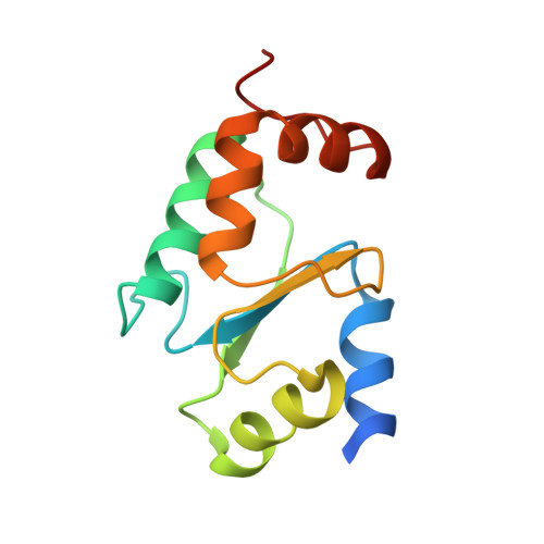
2WUL

CRYSTAL STRUCTURE OF THE HUMAN GLUTAREDOXIN 5 WITH BOUND GLUTATHIONE IN AN FES CLUSTER


Experimental Data Snapshot

  • Method: X-RAY DIFFRACTION
  • Resolution: 2.40 Å
  • R-Value Free: 0.251 
  • R-Value Work: 0.200 
  • R-Value Observed: 0.202 

wwPDB Validation   3D Report Full Report


Ligand Structure Quality Assessment 


This is version 1.4 of the entry. See complete history


Literature

The Crystal Structure of Human Glrx5: Iron Sulphur Cluster Coordination, Tetrameric Assembly and Monomer Activity.

Johansson, C.Roos, A.K.Montano, S.J.Sengupta, R.Filippakopoulos, P.Guo, K.von Delft, F.Holmgren, A.Oppermann, U.Kavanagh, K.L.

(2011) Biochem J 433: 303

  • DOI: https://doi.org/10.1042/BJ20101286
  • Primary Citation of Related Structures:  
    2WUL

  • PubMed Abstract: 

    Human GLRX5 (glutaredoxin 5) is an evolutionarily conserved thiol-disulfide oxidoreductase that has a direct role in the maintenance of normal cytosolic and mitochondrial iron homoeostasis, and its expression affects haem biosynthesis and erythropoiesis. We have crystallized the human GLRX5 bound to two [2Fe-2S] clusters and four GSH molecules. The crystal structure revealed a tetrameric organization with the [2Fe-2S] clusters buried in the interior and shielded from the solvent by the conserved β1-α2 loop, Phe⁶⁹ and the GSH molecules. Each [2Fe-2S] cluster is ligated by the N-terminal activesite cysteine (Cys⁶⁷) thiols contributed by two protomers and two cysteine thiols from two GSH. The two subunits co-ordinating the cluster are in a more extended conformation compared with iron-sulfur-bound human GLRX2, and the intersubunit interactions are more extensive and involve conserved residues among monothiol GLRXs. Gel-filtration chromatography and analytical ultracentrifugation support a tetrameric organization of holo-GLRX5, whereas the apoprotein is monomeric. MS analyses revealed glutathionylation of the cysteine residues in the absence of the [2Fe-2S] cluster, which would protect them from further oxidation and possibly facilitate cluster transfer/acceptance. Apo-GLRX5 reduced glutathione mixed disulfides with a rate 100 times lower than did GLRX2 and was active as a glutathione-dependent electron donor for mammalian ribonucleotide reductase.


  • Organizational Affiliation

    University of Oxford, UK. catrine.johansson@sgc.ox.ac.uk


Macromolecules
Find similar proteins by:  (by identity cutoff)  |  3D Structure
Entity ID: 1
MoleculeChains Sequence LengthOrganismDetailsImage
GLUTAREDOXIN RELATED PROTEIN 5
A, B, C, D
118Homo sapiensMutation(s): 0 
UniProt & NIH Common Fund Data Resources
Find proteins for Q86SX6 (Homo sapiens)
Explore Q86SX6 
Go to UniProtKB:  Q86SX6
PHAROS:  Q86SX6
GTEx:  ENSG00000182512 
Entity Groups  
Sequence Clusters30% Identity50% Identity70% Identity90% Identity95% Identity100% Identity
UniProt GroupQ86SX6
Sequence Annotations
Expand
  • Reference Sequence
Small Molecules
Ligands 4 Unique
IDChains Name / Formula / InChI Key2D Diagram3D Interactions
GSH
Query on GSH

Download Ideal Coordinates CCD File 
E [auth A],
G [auth B],
J [auth C],
L [auth D]
GLUTATHIONE
C10 H17 N3 O6 S
RWSXRVCMGQZWBV-WDSKDSINSA-N
FES
Query on FES

Download Ideal Coordinates CCD File 
F [auth A],
K [auth C]
FE2/S2 (INORGANIC) CLUSTER
Fe2 S2
NIXDOXVAJZFRNF-UHFFFAOYSA-N
PEG
Query on PEG

Download Ideal Coordinates CCD File 
I [auth B]DI(HYDROXYETHYL)ETHER
C4 H10 O3
MTHSVFCYNBDYFN-UHFFFAOYSA-N
CL
Query on CL

Download Ideal Coordinates CCD File 
H [auth B],
M [auth D],
N [auth D],
O [auth D]
CHLORIDE ION
Cl
VEXZGXHMUGYJMC-UHFFFAOYSA-M
Experimental Data & Validation

Experimental Data

  • Method: X-RAY DIFFRACTION
  • Resolution: 2.40 Å
  • R-Value Free: 0.251 
  • R-Value Work: 0.200 
  • R-Value Observed: 0.202 
  • Space Group: P 43 21 2
Unit Cell:
Length ( Å )Angle ( ˚ )
a = 68.995α = 90
b = 68.995β = 90
c = 229.331γ = 90
Software Package:
Software NamePurpose
MOSFLMdata reduction
SCALAdata scaling
SHELXphasing
SHARPphasing
REFMACrefinement

Structure Validation

View Full Validation Report


Ligand Structure Quality Assessment 


Entry History 

Revision History  (Full details and data files)

  • Version 1.0: 2009-10-20
    Type: Initial release
  • Version 1.1: 2011-07-13
    Changes: Advisory, Version format compliance
  • Version 1.2: 2011-12-07
    Changes: Atomic model, Database references, Derived calculations, Non-polymer description
  • Version 1.3: 2018-01-24
    Changes: Database references, Structure summary
  • Version 1.4: 2023-12-20
    Changes: Data collection, Database references, Derived calculations, Other, Refinement description