2Y0G

X-ray structure of Enhanced Green Fluorescent Protein (EGFP)


Experimental Data Snapshot

  • Method: X-RAY DIFFRACTION
  • Resolution: 1.50 Å
  • R-Value Free: 0.188 
  • R-Value Work: 0.136 
  • R-Value Observed: 0.138 

wwPDB Validation   3D Report Full Report


This is version 1.4 of the entry. See complete history


Literature

Stabilizing Role of Glutamic Acid 222 in the Structure of Enhanced Green Fluorescent Protein.

Royant, A.Noirclerc-Savoye, M.

(2011) J Struct Biol 174: 385

  • DOI: https://doi.org/10.1016/j.jsb.2011.02.004
  • Primary Citation of Related Structures:  
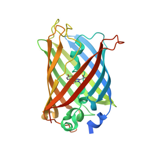
    2Y0G

  • PubMed Abstract: 

    Enhanced Green Fluorescent Protein (EGFP) is a variant of wild-type Green Fluorescent Protein from the jellyfish Aequorea victoria, whose mutations S65T and F64L increase brightness and folding efficiency. EGFP is extensively used in cell biology and biochemistry as a colocalization or expression reporter. Surprisingly, the structure of this very popular protein has not been determined yet. We report here its crystallographic structure at 1.5Å resolution which shows significant differences in the vicinity of residue 64 and of the chromophore. In particular, two conformations are observed for the key residue glutamic acid 222, in apparent contradiction with the single fluorescence lifetime of the protein. We then show that X-ray induced decarboxylation of Glu222 during diffraction data collection results in the disruption of a hydrogen-bond network near the chromophore. Using single-crystal microspectrophotometry, we demonstrate that this correlates with a significant loss of the fluorescence properties. We thus propose a mechanism of bleaching of the protein at low temperature. Taken together, these two sets of results highlight the stabilizing role of Glu222 to the chromophore cavity of EGFP.


  • Organizational Affiliation

    CNRS, Institut de Biologie Structurale Jean-Pierre Ebel, 41 rue Jules Horowitz, 38027 Grenoble, France. antoine.royant@ibs.fr


Macromolecules
Find similar proteins by:  (by identity cutoff)  |  3D Structure
Entity ID: 1
MoleculeChains Sequence LengthOrganismDetailsImage
GREEN FLUORESCENT PROTEIN250Aequorea victoriaMutation(s): 3 
UniProt
Find proteins for P42212 (Aequorea victoria)
Explore P42212 
Go to UniProtKB:  P42212
Entity Groups  
Sequence Clusters30% Identity50% Identity70% Identity90% Identity95% Identity100% Identity
UniProt GroupP42212
Sequence Annotations
Expand
  • Reference Sequence
Small Molecules
Modified Residues  1 Unique
IDChains TypeFormula2D DiagramParent
CRO
Query on CRO
A
L-PEPTIDE LINKINGC15 H17 N3 O5THR, TYR, GLY
Experimental Data & Validation

Experimental Data

  • Method: X-RAY DIFFRACTION
  • Resolution: 1.50 Å
  • R-Value Free: 0.188 
  • R-Value Work: 0.136 
  • R-Value Observed: 0.138 
  • Space Group: P 21 21 21
Unit Cell:
Length ( Å )Angle ( ˚ )
a = 51.047α = 90
b = 62.183β = 90
c = 70.018γ = 90
Software Package:
Software NamePurpose
REFMACrefinement
MOSFLMdata reduction
SCALAdata scaling
MOLREPphasing

Structure Validation

View Full Validation Report


Entry History 

Deposition Data

Revision History  (Full details and data files)

  • Version 1.0: 2011-04-27
    Type: Initial release
  • Version 1.1: 2011-05-08
    Changes: Version format compliance
  • Version 1.2: 2011-07-13
    Changes: Version format compliance
  • Version 1.3: 2019-10-23
    Changes: Data collection, Database references, Derived calculations, Other
  • Version 1.4: 2023-12-20
    Changes: Data collection, Database references, Refinement description