


Structure and Functional Studies on Human Mitochondrial Ferredoxins
Webert, H., Hobler, A., Sheftel, A.D., Molik, S., Maestre-Reyna, M., Essen, L.-O., Vorniscescu, D., Keusgen, M., Hannemann, F., Bernhardt, R., Lill, R.To be published.
Experimental Data Snapshot
wwPDB Validation 3D Report Full Report

Entity ID: 1 | |||||
|---|---|---|---|---|---|
| Molecule | Chains | Sequence Length | Organism | Details | Image |
| ADRENODOXIN-LIKE PROTEIN, MITOCHONDRIAL | 109 | Homo sapiens | Mutation(s): 0 | 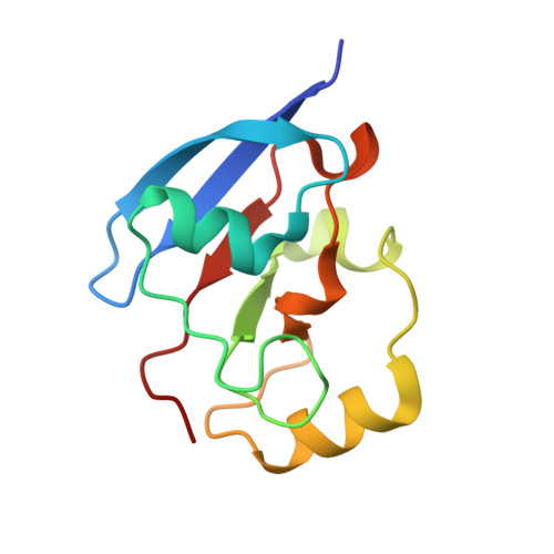 | |
UniProt & NIH Common Fund Data Resources | |||||
Find proteins for Q6P4F2 (Homo sapiens) Explore Q6P4F2 Go to UniProtKB: Q6P4F2 | |||||
PHAROS: Q6P4F2 GTEx: ENSG00000267673 | |||||
Entity Groups | |||||
| Sequence Clusters | 30% Identity50% Identity70% Identity90% Identity95% Identity100% Identity | ||||
| UniProt Group | Q6P4F2 | ||||
Sequence AnnotationsExpand | |||||
| |||||
| Ligands 2 Unique | |||||
|---|---|---|---|---|---|
| ID | Chains | Name / Formula / InChI Key | 2D Diagram | 3D Interactions | |
| FES Query on FES | C [auth A], E [auth B] | FE2/S2 (INORGANIC) CLUSTER Fe2 S2 NIXDOXVAJZFRNF-UHFFFAOYSA-N |  | ||
| SO4 Query on SO4 | D [auth A], F [auth B] | SULFATE ION O4 S QAOWNCQODCNURD-UHFFFAOYSA-L |  | ||
| Length ( Å ) | Angle ( ˚ ) |
|---|---|
| a = 51.38 | α = 90 |
| b = 35.45 | β = 105.13 |
| c = 75.51 | γ = 90 |
| Software Name | Purpose |
|---|---|
| REFMAC | refinement |
| XDS | data reduction |
| SCALA | data scaling |
| SHELXCDE | phasing |
RCSB PDB (citation) is hosted by
RCSB PDB is a member of the

