2YKX | pdb_00002ykx

Structural Determinants of the Beta-Selectivity of a Bacterial Aminotransferase


Experimental Data Snapshot

  • Method: X-RAY DIFFRACTION
  • Resolution: 1.85 Å
  • R-Value Free: 
    0.197 (Depositor), 0.200 (DCC) 
  • R-Value Work: 
    0.176 (Depositor), 0.180 (DCC) 
  • R-Value Observed: 
    0.177 (Depositor) 

Starting Model: other
View more details

wwPDB Validation   3D Report Full Report


Ligand Structure Quality Assessment 

Created with Raphaël 2.3.0Worse 01 BetterLigand structure goodness of fit to experimental dataBest fitted PLPClick on this verticalbar to view details

This is version 1.3 of the entry. See complete history


Literature

Structural Determinants of the Beta-Selectivity of a Bacterial Aminotransferase.

Wybenga, G.G.Crismaru, C.G.Janssen, D.B.Dijkstra, B.W.

(2012) J Biological Chem 287: 28495

  • DOI: https://doi.org/10.1074/jbc.M112.375238
  • Primary Citation of Related Structures:  
    2YKU, 2YKV, 2YKX, 2YKY, 4AO4

  • PubMed Abstract: 

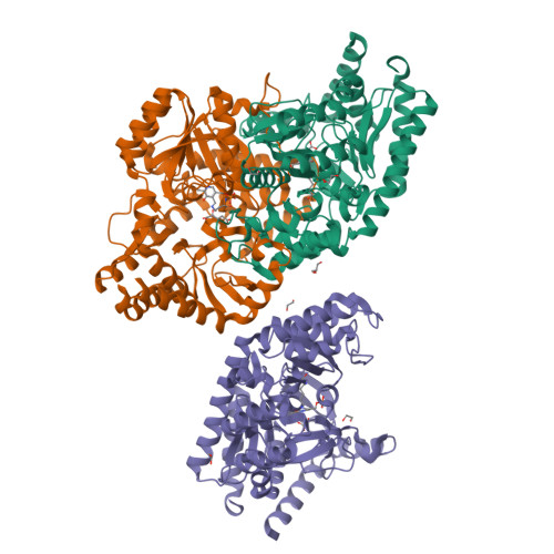
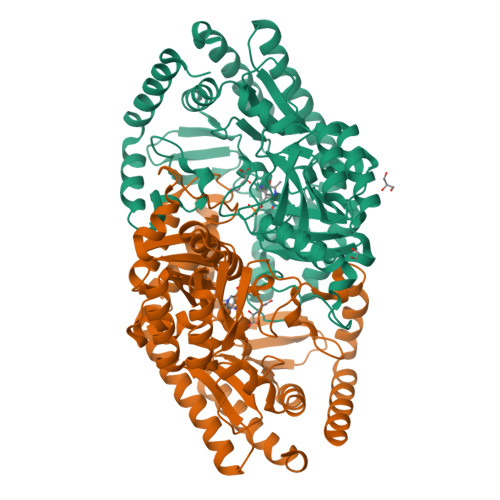
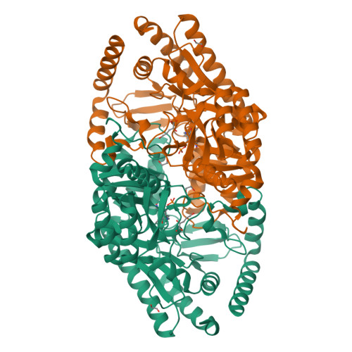
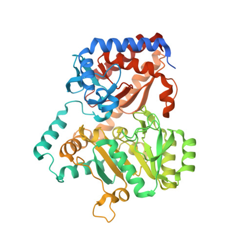
    Chiral β-amino acids occur as constituents of various natural and synthetic compounds with potentially useful bioactivities. The pyridoxal 5'-phosphate (PLP)-dependent S-selective transaminase from Mesorhizobium sp. strain LUK (MesAT) is a fold type I aminotransferase that can be used for the preparation of enantiopure β-Phe and derivatives thereof. Using x-ray crystallography, we solved structures of MesAT in complex with (S)-β-Phe, (R)-3-amino-5-methylhexanoic acid, 2-oxoglutarate, and the inhibitor 2-aminooxyacetic acid, which allowed us to unveil the molecular basis of the amino acid specificity and enantioselectivity of this enzyme. The binding pocket of the side chain of a β-amino acid is located on the 3'-oxygen side of the PLP cofactor. The same binding pocket is utilized by MesAT to bind the α-carboxylate group of an α-amino acid. A β-amino acid thus binds in a reverse orientation in the active site of MesAT compared with an α-amino acid. Such a binding mode has not been reported before for any PLP-dependent aminotransferase and shows that the active site of MesAT has specifically evolved to accommodate both β- and α-amino acids.


  • Organizational Affiliation

    Laboratory of Biophysical Chemistry, University of Groningen, 9747 AG Groningen, The Netherlands.


Macromolecules
Find similar proteins by:  (by identity cutoff)  |  3D Structure
Entity ID: 1
MoleculeChains Sequence LengthOrganismDetailsImage
BETA-TRANSAMINASE
A, B, C
465Mesorhizobium sp. LUKMutation(s): 0 
UniProt
Find proteins for A3EYF7 (Mesorhizobium sp. LUK)
Explore A3EYF7 
Go to UniProtKB:  A3EYF7
Entity Groups  
Sequence Clusters30% Identity50% Identity70% Identity90% Identity95% Identity100% Identity
UniProt GroupA3EYF7
Sequence Annotations
Expand
  • Reference Sequence
Small Molecules
Ligands 4 Unique
IDChains Name / Formula / InChI Key2D Diagram3D Interactions
PLP
Query on PLP

Download Ideal Coordinates CCD File 
D [auth A],
I [auth B],
K [auth C]
PYRIDOXAL-5'-PHOSPHATE
C8 H10 N O6 P
NGVDGCNFYWLIFO-UHFFFAOYSA-N
AKG
Query on AKG

Download Ideal Coordinates CCD File 
E [auth A],
J [auth B],
M [auth C]
2-OXOGLUTARIC ACID
C5 H6 O5
KPGXRSRHYNQIFN-UHFFFAOYSA-N
GOL
Query on GOL

Download Ideal Coordinates CCD File 
F [auth A],
H [auth A]
GLYCEROL
C3 H8 O3
PEDCQBHIVMGVHV-UHFFFAOYSA-N
EDO
Query on EDO

Download Ideal Coordinates CCD File 
G [auth A],
L [auth C],
N [auth C],
O [auth C]
1,2-ETHANEDIOL
C2 H6 O2
LYCAIKOWRPUZTN-UHFFFAOYSA-N
Experimental Data & Validation

Experimental Data

  • Method: X-RAY DIFFRACTION
  • Resolution: 1.85 Å
  • R-Value Free:  0.197 (Depositor), 0.200 (DCC) 
  • R-Value Work:  0.176 (Depositor), 0.180 (DCC) 
  • R-Value Observed: 0.177 (Depositor) 
Space Group: C 1 2 1
Unit Cell:
Length ( Å )Angle ( ˚ )
a = 183.761α = 90
b = 94.422β = 113.65
c = 103.729γ = 90
Software Package:
Software NamePurpose
REFMACrefinement
XDSdata reduction
SCALAdata scaling
MOLREPphasing

Structure Validation

View Full Validation Report


Ligand Structure Quality Assessment 

Created with Raphaël 2.3.0Worse 01 BetterLigand structure goodness of fit to experimental dataBest fitted PLPClick on this verticalbar to view details

Entry History 

Deposition Data

Revision History  (Full details and data files)

  • Version 1.0: 2012-05-30
    Type: Initial release
  • Version 1.1: 2012-07-11
    Changes: Other
  • Version 1.2: 2012-08-29
    Changes: Database references
  • Version 1.3: 2024-05-01
    Changes: Data collection, Database references, Derived calculations, Other, Refinement description