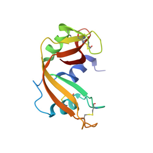
A neutron crystallographic analysis of phosphate-free ribonuclease A at 1.7 A resolution
Yagi, D., Yamada, T., Kurihara, K., Ohnishi, Y., Yamashita, M., Tamada, T., Tanaka, I., Kuroki, R., Niimura, N.(2009) Acta Crystallogr D Biol Crystallogr 65: 892-899
- PubMed: 19690366
- DOI: https://doi.org/10.1107/S0907444909018885
- Primary Citation of Related Structures:
3A1R - PubMed Abstract:
A neutron crystallographic analysis of phosphate-free bovine pancreatic RNase A has been carried out at 1.7 A resolution using the BIX-4 single-crystal diffractometer at the JRR-3 reactor of the Japan Atomic Energy Agency. The high-resolution structural model allowed us to determine that His12 acts mainly as a general base in the catalytic process of RNase A. Numerous other distinctive structural features such as the hydrogen positions of methyl groups, hydroxyl groups, prolines, asparagines and glutamines were also determined at 1.7 A resolution. The protonation and deprotonation states of all of the charged amino-acid residues allowed us to provide a definitive description of the hydrogen-bonding network around the active site and the H atoms of the key His48 residue. Differences in hydrogen-bond strengths for the alpha-helices and beta-sheets were inferred from determination of the hydrogen-bond lengths and the H/D-exchange ratios of the backbone amide H atoms. The correlation between the B factors and hydrogen-bond lengths of the hydration water molecules was also determined.
Organizational Affiliation:
Graduate School of Science and Engineering, Ibaraki University, Hitachi, Japan.


