3A2B

Crystal Structure of Serine Palmitoyltransferase from Sphingobacterium multivorum with substrate L-serine


Experimental Data Snapshot

  • Method: X-RAY DIFFRACTION
  • Resolution: 2.30 Å
  • R-Value Free: 0.270 
  • R-Value Work: 0.211 

wwPDB Validation   3D Report Full Report


Ligand Structure Quality Assessment 


This is version 1.2 of the entry. See complete history


Literature

Structural Insights into the Enzymatic Mechanism of Serine Palmitoyltransferase from Sphingobacterium multivorum

Ikushiro, H.Islam, M.M.Okamoto, A.Hoseki, J.Murakawa, T.Fujii, S.Miyahara, I.Hayashi, H.

(2009) J Biochem 146: 549-562

  • DOI: https://doi.org/10.1093/jb/mvp100
  • Primary Citation of Related Structures:  
    3A2B

  • PubMed Abstract: 

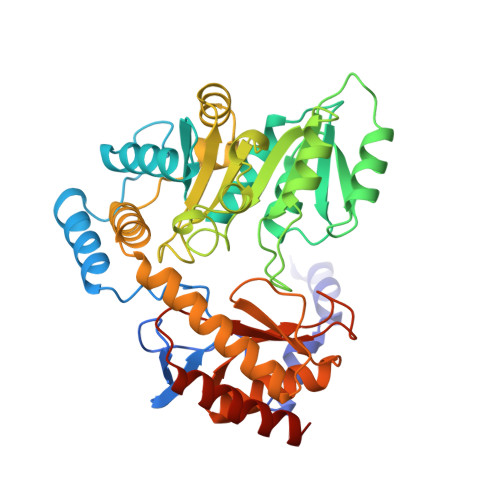
    Serine palmitoyltransferase (SPT) is a key enzyme of sphingolipid biosynthesis and catalyses the pyridoxal 5'-phosphate (PLP)-dependent decarboxylative condensation reaction of l-serine with palmitoyl-CoA to generate 3-ketodihydrosphingosine. The crystal structure of SPT from Sphingobacterium multivorum GTC97 complexed with l-serine was determined at 2.3 A resolution. The electron density map showed the Schiff base formation between l-serine and PLP in the crystal. Because of the hydrogen bond formation with His138, the orientation of the Calpha-H bond of the PLP-l-serine aldimine was not perpendicular to the PLP-Schiff base plane. This conformation is unfavourable for the alpha-proton abstraction by Lys244 and the reaction is expected to stop at the PLP-l-serine aldimine. Structural modelling of the following intermediates indicated that His138 changes its hydrogen bond partner from the carboxyl group of l-serine to the carbonyl group of palmitoyl-CoA upon the binding of palmitoyl-CoA, making the l-serine Calpha-H bond perpendicular to the PLP-Schiff base plane. These crystal and model structures well explained the observations on bacterial SPTs that the alpha-deprotonation of l-serine occurs only in the presence of palmitoyl-CoA. This study provides the structural evidence that directly supports our proposed mechanism of the substrate synergism in the SPT reaction.


  • Organizational Affiliation

    Department of Biochemistry, Osaka Medical College, Takatsuki, Osaka 569-8686, Japan. ikushiro@art.osaka-med.ac.jp


Macromolecules
Find similar proteins by:  (by identity cutoff)  |  3D Structure
Entity ID: 1
MoleculeChains Sequence LengthOrganismDetailsImage
Serine palmitoyltransferase398Sphingobacterium multivorumMutation(s): 0 
Gene Names: spt
EC: 2.3.1.50
UniProt
Find proteins for A7BFV6 (Sphingobacterium multivorum)
Explore A7BFV6 
Go to UniProtKB:  A7BFV6
Entity Groups  
Sequence Clusters30% Identity50% Identity70% Identity90% Identity95% Identity100% Identity
UniProt GroupA7BFV6
Sequence Annotations
Expand
  • Reference Sequence
Small Molecules
Ligands 2 Unique
IDChains Name / Formula / InChI Key2D Diagram3D Interactions
PLP
Query on PLP

Download Ideal Coordinates CCD File 
C [auth A]PYRIDOXAL-5'-PHOSPHATE
C8 H10 N O6 P
NGVDGCNFYWLIFO-UHFFFAOYSA-N
SER
Query on SER

Download Ideal Coordinates CCD File 
B [auth A]SERINE
C3 H7 N O3
MTCFGRXMJLQNBG-REOHCLBHSA-N
Experimental Data & Validation

Experimental Data

  • Method: X-RAY DIFFRACTION
  • Resolution: 2.30 Å
  • R-Value Free: 0.270 
  • R-Value Work: 0.211 
  • Space Group: P 41 21 2
Unit Cell:
Length ( Å )Angle ( ˚ )
a = 61.67α = 90
b = 61.67β = 90
c = 207.76γ = 90
Software Package:
Software NamePurpose
MOLREPphasing
CNSrefinement
HKL-2000data collection
HKL-2000data reduction
SCALEPACKdata scaling

Structure Validation

View Full Validation Report


Ligand Structure Quality Assessment 


Entry History 

Deposition Data

Revision History  (Full details and data files)

  • Version 1.0: 2009-07-14
    Type: Initial release
  • Version 1.1: 2011-07-13
    Changes: Non-polymer description, Version format compliance
  • Version 1.2: 2023-11-01
    Changes: Data collection, Database references, Derived calculations, Refinement description