3BG8

Crystal structure of Factor XIa in complex with Clavatadine A


Experimental Data Snapshot

  • Method: X-RAY DIFFRACTION
  • Resolution: 1.60 Å
  • R-Value Free: 0.200 
  • R-Value Work: 0.178 
  • R-Value Observed: 0.179 

wwPDB Validation   3D Report Full Report


Ligand Structure Quality Assessment 


This is version 2.0 of the entry. See complete history


Literature

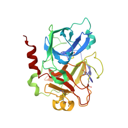
Clavatadine A, a natural product with selective recognition and irreversible inhibition of factor XIa.

Buchanan, M.S.Carroll, A.R.Wessling, D.Jobling, M.Avery, V.M.Davis, R.A.Feng, Y.Xue, Y.Oster, L.Fex, T.Deinum, J.Hooper, J.N.Quinn, R.J.

(2008) J Med Chem 51: 3583-3587

  • DOI: https://doi.org/10.1021/jm800314b
  • Primary Citation of Related Structures:  
    3BG8

  • PubMed Abstract: 

    Bioassay-guided fractionation of a CH2Cl2/MeOH extract of the sponge Suberea clavata using the serine protease factor XIa to detect antithrombotic activity led to the isolation of the new marine natural products, clavatadines A and B. Clavatadines A and B inhibited factor XIa with IC50's of 1.3 and 27 microM, respectively. A crystal structure of protein-inhibitor (clavatadine A) complex was obtained and revealed interesting selective binding and irreversible inhibition of factor XIa. The cocrystal structure provides guidance for the design and synthesis of future factor XIa inhibitors as antithrombotic agents.


  • Organizational Affiliation

    Eskitis Institute, Griffith University, Brisbane, Queensland 4111, Australia.


Macromolecules
Find similar proteins by:  (by identity cutoff)  |  3D Structure
Entity ID: 1
MoleculeChains Sequence LengthOrganismDetailsImage
Coagulation factor XIa light chain238Homo sapiensMutation(s): 0 
Gene Names: F11
EC: 3.4.21.27
UniProt & NIH Common Fund Data Resources
Find proteins for P03951 (Homo sapiens)
Explore P03951 
Go to UniProtKB:  P03951
PHAROS:  P03951
GTEx:  ENSG00000088926 
Entity Groups  
Sequence Clusters30% Identity50% Identity70% Identity90% Identity95% Identity100% Identity
UniProt GroupP03951
Sequence Annotations
Expand
  • Reference Sequence
Experimental Data & Validation

Experimental Data

  • Method: X-RAY DIFFRACTION
  • Resolution: 1.60 Å
  • R-Value Free: 0.200 
  • R-Value Work: 0.178 
  • R-Value Observed: 0.179 
  • Space Group: I 2 3
Unit Cell:
Length ( Å )Angle ( ˚ )
a = 120.716α = 90
b = 120.716β = 90
c = 120.716γ = 90
Software Package:
Software NamePurpose
REFMACrefinement
ADSCdata collection
MOSFLMdata reduction
SCALAdata scaling
MOLREPphasing

Structure Validation

View Full Validation Report


Ligand Structure Quality Assessment 


Entry History 

Deposition Data

Revision History  (Full details and data files)

  • Version 1.0: 2008-12-09
    Type: Initial release
  • Version 1.1: 2011-07-13
    Changes: Version format compliance
  • Version 2.0: 2021-10-13
    Changes: Advisory, Atomic model, Data collection, Database references, Derived calculations, Non-polymer description, Structure summary