3C31

Crystal structure of GluR5 ligand-binding core in complex with lithium at 1.49 Angstrom resolution


Experimental Data Snapshot

  • Method: X-RAY DIFFRACTION
  • Resolution: 1.49 Å
  • R-Value Free: 0.190 
  • R-Value Work: 0.165 
  • R-Value Observed: 0.167 

wwPDB Validation   3D Report Full Report


Ligand Structure Quality Assessment 


This is version 1.3 of the entry. See complete history


Literature

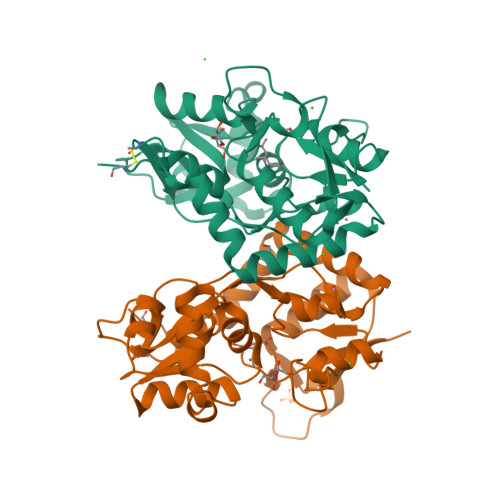
Molecular basis of kainate receptor modulation by sodium.

Plested, A.J.Vijayan, R.Biggin, P.C.Mayer, M.L.

(2008) Neuron 58: 720-735

  • DOI: https://doi.org/10.1016/j.neuron.2008.04.001
  • Primary Citation of Related Structures:  
    3C31, 3C32, 3C33, 3C34, 3C35, 3C36

  • PubMed Abstract: 

    Membrane proteins function in a polarized ionic environment with sodium-rich extracellular and potassium-rich intracellular solutions. Glutamate receptors that mediate excitatory synaptic transmission in the brain show unusual sensitivity to external ions, resulting in an apparent requirement for sodium in order for glutamate to activate kainate receptors. Here, we solve the structure of the Na(+)-binding sites and determine the mechanism by which allosteric anions and cations regulate ligand-binding dimer stability, and hence the rate of desensitization and receptor availability for gating by glutamate. We establish a stoichiometry for binding of 2 Na(+) to 1 Cl(-) and show that allosteric anions and cations bind at physically discrete sites with strong electric fields, that the binding sites are not saturated in CSF, and that the requirement of kainate receptors for Na(+) occurs simply because other cations bind with lower affinity and have lower efficacy compared to Na(+).


  • Organizational Affiliation

    Laboratory of Cellular and Molecular Neurophysiology, Porter Neuroscience Research Center, NICHD, NIH, DHHS, Bethesda, MD 20892, USA.


Macromolecules
Find similar proteins by:  (by identity cutoff)  |  3D Structure
Entity ID: 1
MoleculeChains Sequence LengthOrganismDetailsImage
GLUTAMATE RECEPTOR, IONOTROPIC KAINATE 1
A, B
258Rattus norvegicusMutation(s): 0 
Gene Names: GRIK1GLUR5
UniProt
Find proteins for P22756 (Rattus norvegicus)
Explore P22756 
Go to UniProtKB:  P22756
Entity Groups  
Sequence Clusters30% Identity50% Identity70% Identity90% Identity95% Identity100% Identity
UniProt GroupP22756
Sequence Annotations
Expand
  • Reference Sequence
Small Molecules
Ligands 5 Unique
IDChains Name / Formula / InChI Key2D Diagram3D Interactions
KAI
Query on KAI

Download Ideal Coordinates CCD File 
G [auth A],
N [auth B]
3-(CARBOXYMETHYL)-4-ISOPROPENYLPROLINE
C10 H15 N O4
VLSMHEGGTFMBBZ-OOZYFLPDSA-N
SO4
Query on SO4

Download Ideal Coordinates CCD File 
M [auth B]SULFATE ION
O4 S
QAOWNCQODCNURD-UHFFFAOYSA-L
GOL
Query on GOL

Download Ideal Coordinates CCD File 
H [auth A],
I [auth A],
J [auth A],
O [auth B]
GLYCEROL
C3 H8 O3
PEDCQBHIVMGVHV-UHFFFAOYSA-N
CL
Query on CL

Download Ideal Coordinates CCD File 
D [auth A],
E [auth A],
F [auth A],
L [auth B]
CHLORIDE ION
Cl
VEXZGXHMUGYJMC-UHFFFAOYSA-M
LI
Query on LI

Download Ideal Coordinates CCD File 
C [auth A],
K [auth B]
LITHIUM ION
Li
HBBGRARXTFLTSG-UHFFFAOYSA-N
Binding Affinity Annotations 
IDSourceBinding Affinity
KAI BindingDB:  3C31 Ki: min: 1.41, max: 177 (nM) from 7 assay(s)
Kd: 39 (nM) from 1 assay(s)
IC50: 77 (nM) from 1 assay(s)
EC50: min: 1800, max: 3.70e+4 (nM) from 4 assay(s)
Experimental Data & Validation

Experimental Data

  • Method: X-RAY DIFFRACTION
  • Resolution: 1.49 Å
  • R-Value Free: 0.190 
  • R-Value Work: 0.165 
  • R-Value Observed: 0.167 
  • Space Group: P 41 21 2
Unit Cell:
Length ( Å )Angle ( ˚ )
a = 70.529α = 90
b = 70.529β = 90
c = 234.128γ = 90
Software Package:
Software NamePurpose
REFMACrefinement
HKL-2000data collection
HKL-2000data reduction
HKL-2000data scaling
PHASERphasing

Structure Validation

View Full Validation Report


Ligand Structure Quality Assessment 


Entry History 

Deposition Data

  • Released Date: 2008-06-17 
  • Deposition Author(s): Mayer, M.L.

Revision History  (Full details and data files)

  • Version 1.0: 2008-06-17
    Type: Initial release
  • Version 1.1: 2011-07-13
    Changes: Advisory, Version format compliance
  • Version 1.2: 2017-08-02
    Changes: Source and taxonomy
  • Version 1.3: 2023-08-30
    Changes: Data collection, Database references, Derived calculations, Refinement description