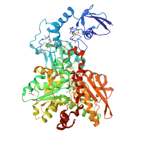
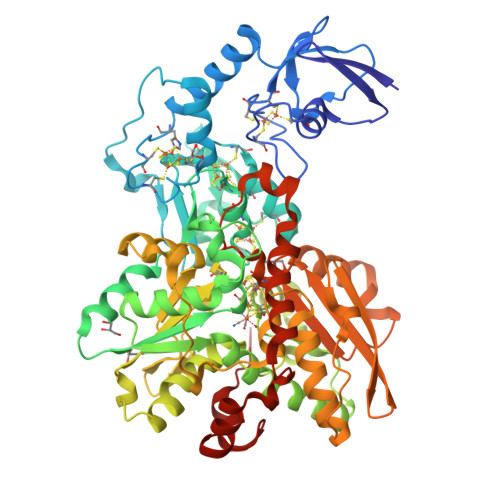
3C8Y | pdb_00003c8y

1.39 Angstrom crystal structure of Fe-only hydrogenase


Experimental Data Snapshot

  • Method: X-RAY DIFFRACTION
  • Resolution: 1.39 Å
  • R-Value Free: 
    0.194 (Depositor) 
  • R-Value Work: 
    0.132 (Depositor), 0.140 (DCC) 

Starting Model: experimental
View more details

wwPDB Validation   3D Report Full Report


Ligand Structure Quality Assessment 

Created with Raphaël 2.3.0Worse 01 BetterLigand structure goodness of fit to experimental dataBest fitted HCNClick on this verticalbar to view details

This is version 1.2 of the entry. See complete history


Literature

Dithiomethylether as a ligand in the hydrogenase h-cluster.

Pandey, A.S.Harris, T.V.Giles, L.J.Peters, J.W.Szilagyi, R.K.

(2008) J Am Chem Soc 130: 4533-4540

  • DOI: https://doi.org/10.1021/ja711187e
  • Primary Citation of Related Structures:  
    3C8Y

  • PubMed Abstract: 

    An X-ray crystallographic refinement of the H-cluster of [FeFe]-hydrogenase from Clostridium pasteurianum has been carried out to close-to atomic resolution and is the highest resolution [FeFe]-hydrogenase presented to date. The 1.39 A, anisotropically refined [FeFe]-hydrogenase structure provides a basis for examining the outstanding issue of the composition of the unique nonprotein dithiolate ligand of the H-cluster. In addition to influencing the electronic structure of the H-cluster, the composition of the ligand has mechanistic implications due to the potential of the bridge-head gamma-group participating in proton transfer during catalysis. In this work, sequential density functional theory optimizations of the dithiolate ligand embedded in a 3.5-3.9 A protein environment provide an unbiased approach to examining the most likely composition of the ligand. Structural, conformational, and energetic considerations indicate a preference for dithiomethylether as an H-cluster ligand and strongly disfavor the dithiomethylammonium as a catalytic base for hydrogen production.


  • Organizational Affiliation

    Department of Chemistry and Biochemistry and Astrobiology Biogeocatalysis Research Center, Montana State University, Bozeman, Montana 59717, USA.


Macromolecules
Find similar proteins by:  (by identity cutoff)  |  3D Structure
Entity ID: 1
MoleculeChains Sequence LengthOrganismDetailsImage
Iron hydrogenase 1574Clostridium pasteurianumMutation(s): 0 
EC: 1.12.7.2
UniProt
Find proteins for P29166 (Clostridium pasteurianum)
Explore P29166 
Go to UniProtKB:  P29166
Entity Groups  
Sequence Clusters30% Identity50% Identity70% Identity90% Identity95% Identity100% Identity
UniProt GroupP29166
Sequence Annotations
Expand
  • Reference Sequence
Small Molecules
Ligands 4 Unique
IDChains Name / Formula / InChI Key2D Diagram3D Interactions
HCN
Query on HCN

Download Ideal Coordinates CCD File 
B [auth A]2 IRON/2 SULFUR/3 CARBONYL/2 CYANIDE/WATER/METHYLETHER CLUSTER
C7 H10 Fe2 N2 O5 S2
GIQCNDUJZDHMQH-UHFFFAOYSA-N
SF4
Query on SF4

Download Ideal Coordinates CCD File 
C [auth A],
D [auth A],
E [auth A],
F [auth A]
IRON/SULFUR CLUSTER
Fe4 S4
LJBDFODJNLIPKO-UHFFFAOYSA-N
FES
Query on FES

Download Ideal Coordinates CCD File 
G [auth A]FE2/S2 (INORGANIC) CLUSTER
Fe2 S2
NIXDOXVAJZFRNF-UHFFFAOYSA-N
GOL
Query on GOL

Download Ideal Coordinates CCD File 
H [auth A],
I [auth A],
J [auth A]
GLYCEROL
C3 H8 O3
PEDCQBHIVMGVHV-UHFFFAOYSA-N
Experimental Data & Validation

Experimental Data

  • Method: X-RAY DIFFRACTION
  • Resolution: 1.39 Å
  • R-Value Free:  0.194 (Depositor) 
  • R-Value Work:  0.132 (Depositor), 0.140 (DCC) 
Space Group: P 42 21 2
Unit Cell:
Length ( Å )Angle ( ˚ )
a = 110.75α = 90
b = 110.75β = 90
c = 103.54γ = 90
Software Package:
Software NamePurpose
MOSFLMdata reduction
SCALAdata scaling
MOLREPphasing
SHELXrefinement
PDB_EXTRACTdata extraction
SHELXL-97refinement

Structure Validation

View Full Validation Report


Ligand Structure Quality Assessment 

Created with Raphaël 2.3.0Worse 01 BetterLigand structure goodness of fit to experimental dataBest fitted HCNClick on this verticalbar to view details

Entry History 

Deposition Data

Revision History  (Full details and data files)

  • Version 1.0: 2008-04-22
    Type: Initial release
  • Version 1.1: 2011-07-13
    Changes: Non-polymer description, Version format compliance
  • Version 1.2: 2023-08-30
    Changes: Data collection, Database references, Derived calculations, Refinement description