3CS8

Structural and Biochemical Basis for the Binding Selectivity of PPARg to PGC-1a


Experimental Data Snapshot

  • Method: X-RAY DIFFRACTION
  • Resolution: 2.30 Å
  • R-Value Free: 0.282 
  • R-Value Work: 0.224 
  • R-Value Observed: 0.227 

wwPDB Validation   3D Report Full Report


Ligand Structure Quality Assessment 


This is version 1.2 of the entry. See complete history


Literature

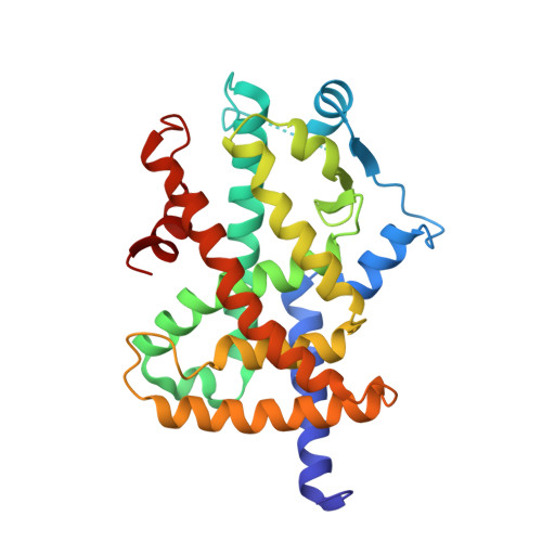
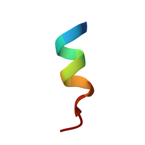
Structural and biochemical basis for the binding selectivity of peroxisome proliferator-activated receptor gamma to PGC-1alpha.

Li, Y.Kovach, A.Suino-Powell, K.Martynowski, D.Xu, H.E.

(2008) J Biol Chem 283: 19132-19139

  • DOI: https://doi.org/10.1074/jbc.M802040200
  • Primary Citation of Related Structures:  
    3CS8

  • PubMed Abstract: 

    The functional interaction between the peroxisome proliferator-activated receptor gamma (PPARgamma) and its coactivator PGC-1alpha is crucial for the normal physiology of PPARgamma and its pharmacological response to antidiabetic treatment with rosiglitazone. Here we report the crystal structure of the PPARgamma ligand-binding domain bound to rosiglitazone and to a large PGC-1alpha fragment that contains two LXXLL-related motifs. The structure reveals critical contacts mediated through the first LXXLL motif of PGC-1alpha and the PPARgamma coactivator binding site. Through a combination of biochemical and structural studies, we demonstrate that the first LXXLL motif is the most potent among all nuclear receptor coactivator motifs tested, and only this motif of the two LXXLL-related motifs in PGC-1alpha is capable of binding to PPARgamma. Our studies reveal that the strong interaction of PGC-1alpha and PPARgamma is mediated through both hydrophobic and specific polar interactions. Mutations within the context of the full-length PGC-1alpha indicate that the first PGC-1alpha motif is necessary and sufficient for PGC-1alpha to coactivate PPARgamma in the presence or absence of rosiglitazone. These results provide a molecular basis for specific recruitment and functional interplay between PPARgamma and PGC-1alpha in glucose homeostasis and adipocyte differentiation.


  • Organizational Affiliation

    Laboratory of Structural Sciences, Van Andel Research Institute, Grand Rapids, Michigan 49503, USA. yol21@pitt.edu


Macromolecules
Find similar proteins by:  (by identity cutoff)  |  3D Structure
Entity ID: 1
MoleculeChains Sequence LengthOrganismDetailsImage
Peroxisome proliferator-activated receptor gamma275Homo sapiensMutation(s): 0 
Gene Names: PPARGC1ALEM6PGC1PGC1APPARGC1
UniProt & NIH Common Fund Data Resources
Find proteins for P37231 (Homo sapiens)
Explore P37231 
Go to UniProtKB:  P37231
PHAROS:  P37231
GTEx:  ENSG00000132170 
Entity Groups  
Sequence Clusters30% Identity50% Identity70% Identity90% Identity95% Identity100% Identity
UniProt GroupP37231
Sequence Annotations
Expand
  • Reference Sequence

Find similar proteins by:  Sequence   |   3D Structure  

Entity ID: 2
MoleculeChains Sequence LengthOrganismDetailsImage
PGC-1alfa peptide12N/AMutation(s): 0 
UniProt & NIH Common Fund Data Resources
Find proteins for Q9UBK2 (Homo sapiens)
Explore Q9UBK2 
Go to UniProtKB:  Q9UBK2
PHAROS:  Q9UBK2
GTEx:  ENSG00000109819 
Entity Groups  
Sequence Clusters30% Identity50% Identity70% Identity90% Identity95% Identity100% Identity
UniProt GroupQ9UBK2
Sequence Annotations
Expand
  • Reference Sequence
Small Molecules
Binding Affinity Annotations 
IDSourceBinding Affinity
BRL BindingDB:  3CS8 Ki: min: 8, max: 59 (nM) from 5 assay(s)
Kd: min: 0.14, max: 48 (nM) from 3 assay(s)
IC50: min: 1, max: 298 (nM) from 10 assay(s)
EC50: min: 1, max: 1600 (nM) from 35 assay(s)
Experimental Data & Validation

Experimental Data

  • Method: X-RAY DIFFRACTION
  • Resolution: 2.30 Å
  • R-Value Free: 0.282 
  • R-Value Work: 0.224 
  • R-Value Observed: 0.227 
  • Space Group: C 1 2 1
Unit Cell:
Length ( Å )Angle ( ˚ )
a = 95.111α = 90
b = 53.887β = 104.83
c = 64.889γ = 90
Software Package:
Software NamePurpose
REFMACrefinement
HKL-2000data collection
DENZOdata reduction
SCALEPACKdata scaling
AMoREphasing

Structure Validation

View Full Validation Report


Ligand Structure Quality Assessment 


Entry History 

Deposition Data

Revision History  (Full details and data files)

  • Version 1.0: 2008-06-03
    Type: Initial release
  • Version 1.1: 2011-07-13
    Changes: Version format compliance
  • Version 1.2: 2024-02-21
    Changes: Data collection, Database references, Derived calculations