3FCI

Complex of UNG2 and a fragment-based designed inhibitor


Experimental Data Snapshot

  • Method: X-RAY DIFFRACTION
  • Resolution: 1.27 Å
  • R-Value Free: 0.207 
  • R-Value Work: 0.174 
  • R-Value Observed: 0.176 

wwPDB Validation   3D Report Full Report


Ligand Structure Quality Assessment 


This is version 1.3 of the entry. See complete history


Literature

Impact of linker strain and flexibility in the design of a fragment-based inhibitor

Chung, S.Parker, J.B.Bianchet, M.Amzel, L.M.Stivers, J.T.

(2009) Nat Chem Biol 5: 407-413

  • DOI: https://doi.org/10.1038/nchembio.163
  • Primary Citation of Related Structures:  
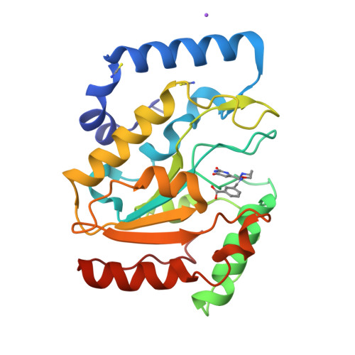
    3FCF, 3FCI, 3FCK, 3FCL

  • PubMed Abstract: 

    The linking together of molecular fragments that bind to adjacent sites on an enzyme can lead to high-affinity inhibitors. Ideally, this strategy would use linkers that do not perturb the optimal binding geometries of the fragments and do not have excessive conformational flexibility that would increase the entropic penalty of binding. In reality, these aims are seldom realized owing to limitations in linker chemistry. Here we systematically explore the energetic and structural effects of rigid and flexible linkers on the binding of a fragment-based inhibitor of human uracil DNA glycosylase. Analysis of the free energies of binding in combination with cocrystal structures shows that the flexibility and strain of a given linker can have a substantial impact on binding affinity even when the binding fragments are optimally positioned. Such effects are not apparent from inspection of structures and underscore the importance of linker optimization in fragment-based drug discovery efforts.


  • Organizational Affiliation

    Department of Pharmacology and Molecular Sciences, Johns Hopkins University School of Medicine, Baltimore, Maryland, USA.


Macromolecules
Find similar proteins by:  (by identity cutoff)  |  3D Structure
Entity ID: 1
MoleculeChains Sequence LengthOrganismDetailsImage
Uracil-DNA glycosylase223Homo sapiensMutation(s): 0 
Gene Names: UNGDGUUNG1UNG15
EC: 3.2.2
UniProt & NIH Common Fund Data Resources
Find proteins for P13051 (Homo sapiens)
Explore P13051 
Go to UniProtKB:  P13051
PHAROS:  P13051
GTEx:  ENSG00000076248 
Entity Groups  
Sequence Clusters30% Identity50% Identity70% Identity90% Identity95% Identity100% Identity
UniProt GroupP13051
Sequence Annotations
Expand
  • Reference Sequence
Small Molecules
Binding Affinity Annotations 
IDSourceBinding Affinity
3FI Binding MOAD:  3FCI IC50: 1300 (nM) from 1 assay(s)
BindingDB:  3FCI IC50: 1300 (nM) from 1 assay(s)
PDBBind:  3FCI IC50: 1300 (nM) from 1 assay(s)
Experimental Data & Validation

Experimental Data

  • Method: X-RAY DIFFRACTION
  • Resolution: 1.27 Å
  • R-Value Free: 0.207 
  • R-Value Work: 0.174 
  • R-Value Observed: 0.176 
  • Space Group: P 21 21 21
Unit Cell:
Length ( Å )Angle ( ˚ )
a = 43.014α = 90
b = 68.476β = 90
c = 69.674γ = 90
Software Package:
Software NamePurpose
DENZOdata reduction
SCALEPACKdata scaling
REFMACrefinement
PDB_EXTRACTdata extraction
ADSCdata collection
HKL-2000data reduction
HKL-2000data scaling
MOLREPphasing

Structure Validation

View Full Validation Report


Ligand Structure Quality Assessment 


Entry History 

Deposition Data

Revision History  (Full details and data files)

  • Version 1.0: 2009-04-28
    Type: Initial release
  • Version 1.1: 2011-07-13
    Changes: Version format compliance
  • Version 1.2: 2017-11-01
    Changes: Refinement description
  • Version 1.3: 2023-12-27
    Changes: Data collection, Database references, Derived calculations