


3IAI
Crystal structure of the catalytic domain of the tumor-associated human carbonic anhydrase IX
- PDB DOI: https://doi.org/10.2210/pdb3IAI/pdb
- Classification: LYASE
- Organism(s): Homo sapiens
- Expression System: Spodoptera frugiperda
- Mutation(s): Yes
- Deposited: 2009-07-14 Released: 2009-09-08
Experimental Data Snapshot
- Method: X-RAY DIFFRACTION
- Resolution: 2.20 Å
- R-Value Free: 0.181
- R-Value Work: 0.157

This is version 2.2 of the entry. See complete history.

Macromolecules
Find similar proteins by:
(by identity cutoff) | 3D Structure
Entity ID: 1 | |||||
|---|---|---|---|---|---|
| Molecule | Chains | Sequence Length | Organism | Details | Image |
| Carbonic anhydrase 9 | 257 | Homo sapiens | Mutation(s): 1 Gene Names: CA9, G250, MN EC: 4.2.1.1 | 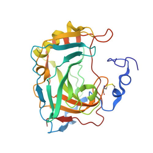 | |
UniProt & NIH Common Fund Data Resources | |||||
Find proteins for Q16790 (Homo sapiens) Explore Q16790 Go to UniProtKB: Q16790 | |||||
PHAROS: Q16790 GTEx: ENSG00000107159 | |||||
Entity Groups | |||||
| Sequence Clusters | 30% Identity50% Identity70% Identity90% Identity95% Identity100% Identity | ||||
| UniProt Group | Q16790 | ||||
Sequence AnnotationsExpand | |||||
| |||||
Oligosaccharides
Entity ID: 2 | |||||
|---|---|---|---|---|---|
| Molecule | Chains | Length | 2D Diagram | Glycosylation | 3D Interactions |
| alpha-D-mannopyranose-(1-3)-[alpha-D-mannopyranose-(1-6)]beta-D-mannopyranose-(1-4)-2-acetamido-2-deoxy-beta-D-glucopyranose-(1-4)-2-acetamido-2-deoxy-beta-D-glucopyranose | E, F, G, H | 5 |  | N-Glycosylation | |
Glycosylation Resources | |||||
GlyTouCan: G22768VO GlyCosmos: G22768VO GlyGen: G22768VO | |||||
Small Molecules
| Ligands 5 Unique | |||||
|---|---|---|---|---|---|
| ID | Chains | Name / Formula / InChI Key | 2D Diagram | 3D Interactions | |
| AZM Query on AZM | FA [auth C], J [auth A], OA [auth D], V [auth B] | 5-ACETAMIDO-1,3,4-THIADIAZOLE-2-SULFONAMIDE C4 H6 N4 O3 S2 BZKPWHYZMXOIDC-UHFFFAOYSA-N |  | ||
| TRS Query on TRS | CA [auth B], LA [auth C], R [auth A], WA [auth D] | 2-AMINO-2-HYDROXYMETHYL-PROPANE-1,3-DIOL C4 H12 N O3 LENZDBCJOHFCAS-UHFFFAOYSA-O |  | ||
| PO4 Query on PO4 | DA [auth B], MA [auth C], S [auth A], T [auth A], XA [auth D] | PHOSPHATE ION O4 P NBIIXXVUZAFLBC-UHFFFAOYSA-K |  | ||
| GOL Query on GOL | AA [auth B] BA [auth B] GA [auth C] HA [auth C] IA [auth C] | GLYCEROL C3 H8 O3 PEDCQBHIVMGVHV-UHFFFAOYSA-N |  | ||
| ZN Query on ZN | EA [auth C], I [auth A], NA [auth D], U [auth B] | ZINC ION Zn PTFCDOFLOPIGGS-UHFFFAOYSA-N |  | ||
Experimental Data & Validation
Experimental Data
- Method: X-RAY DIFFRACTION
- Resolution: 2.20 Å
- R-Value Free: 0.181
- R-Value Work: 0.157
- Space Group: P 61
Unit Cell:
| Length ( Å ) | Angle ( ˚ ) |
|---|---|
| a = 144.18 | α = 90 |
| b = 144.18 | β = 90 |
| c = 208.89 | γ = 120 |
| Software Name | Purpose |
|---|---|
| AMoRE | phasing |
| CNS | refinement |
| HKL-2000 | data reduction |
| HKL-2000 | data scaling |
Entry History
Deposition Data
- Released Date: 2009-09-08 Deposition Author(s): Alterio, V., Di Fiore, A., De Simone, G.
Revision History (Full details and data files)
- Version 1.0: 2009-09-08
Type: Initial release - Version 1.1: 2011-07-13
Changes: Non-polymer description, Version format compliance - Version 2.0: 2020-07-29
Type: Remediation
Reason: Carbohydrate remediation
Changes: Atomic model, Data collection, Database references, Derived calculations, Structure summary - Version 2.1: 2021-10-13
Changes: Database references, Structure summary - Version 2.2: 2023-09-06
Changes: Data collection, Refinement description
