3J5Q

Structure of TRPV1 ion channel in complex with DkTx and RTX determined by single particle electron cryo-microscopy


Experimental Data Snapshot

  • Method: ELECTRON MICROSCOPY
  • Resolution: 3.80 Å
  • Aggregation State: PARTICLE 
  • Reconstruction Method: SINGLE PARTICLE 

wwPDB Validation   3D Report Full Report


This is version 2.0 of the entry. See complete history


Literature

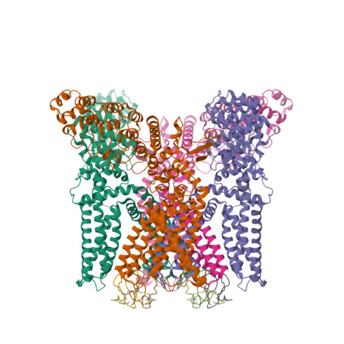
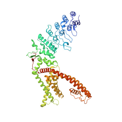
TRPV1 structures in distinct conformations reveal activation mechanisms.

Cao, E.Liao, M.Cheng, Y.Julius, D.

(2013) Nature 504: 113-118

  • DOI: https://doi.org/10.1038/nature12823
  • Primary Citation of Related Structures:  
    3J5Q, 3J5R

  • PubMed Abstract: 

    Transient receptor potential (TRP) channels are polymodal signal detectors that respond to a wide range of physical and chemical stimuli. Elucidating how these channels integrate and convert physiological signals into channel opening is essential to understanding how they regulate cell excitability under normal and pathophysiological conditions. Here we exploit pharmacological probes (a peptide toxin and small vanilloid agonists) to determine structures of two activated states of the capsaicin receptor, TRPV1. A domain (consisting of transmembrane segments 1-4) that moves during activation of voltage-gated channels remains stationary in TRPV1, highlighting differences in gating mechanisms for these structurally related channel superfamilies. TRPV1 opening is associated with major structural rearrangements in the outer pore, including the pore helix and selectivity filter, as well as pronounced dilation of a hydrophobic constriction at the lower gate, suggesting a dual gating mechanism. Allosteric coupling between upper and lower gates may account for rich physiological modulation exhibited by TRPV1 and other TRP channels.


  • Organizational Affiliation

    1] Department of Physiology, University of California, San Francisco, California 94158-2517, USA [2].


Macromolecules
Find similar proteins by:  (by identity cutoff)  |  3D Structure
Entity ID: 1
MoleculeChains Sequence LengthOrganismDetailsImage
Transient receptor potential cation channel subfamily V member 1A [auth D],
B,
C [auth E],
D [auth G]
628Rattus norvegicusMutation(s): 0 
Gene Names: Trpv1Vr1Vr1l
Membrane Entity: Yes 
UniProt
Find proteins for O35433 (Rattus norvegicus)
Explore O35433 
Go to UniProtKB:  O35433
Entity Groups  
Sequence Clusters30% Identity50% Identity70% Identity90% Identity95% Identity100% Identity
UniProt GroupO35433
Sequence Annotations
Expand
  • Reference Sequence
Find similar proteins by:  (by identity cutoff)  |  3D Structure
Entity ID: 2
MoleculeChains Sequence LengthOrganismDetailsImage
Kappa-theraphotoxin-Cg1a 1E [auth A],
F [auth C],
G [auth F],
H
31Chilobrachys guangxiensisMutation(s): 0 
Membrane Entity: Yes 
UniProt
Find proteins for P0C247 (Chilobrachys guangxiensis)
Explore P0C247 
Go to UniProtKB:  P0C247
Entity Groups  
Sequence Clusters30% Identity50% Identity70% Identity90% Identity95% Identity100% Identity
UniProt GroupP0C247
Sequence Annotations
Expand
  • Reference Sequence
Experimental Data & Validation

Experimental Data

  • Method: ELECTRON MICROSCOPY
  • Resolution: 3.80 Å
  • Aggregation State: PARTICLE 
  • Reconstruction Method: SINGLE PARTICLE 
EM Software:
TaskSoftware PackageVersion
RECONSTRUCTIONRELION

Structure Validation

View Full Validation Report


Entry History 

Deposition Data

Revision History  (Full details and data files)

  • Version 1.0: 2013-12-04
    Type: Initial release
  • Version 1.1: 2013-12-11
    Changes: Database references
  • Version 1.2: 2013-12-18
    Changes: Database references
  • Version 1.3: 2018-07-18
    Changes: Data collection
  • Version 2.0: 2022-11-02
    Changes: Advisory, Atomic model, Data collection, Database references, Derived calculations, Polymer sequence, Source and taxonomy, Structure summary