3MAX

Crystal Structure of Human HDAC2 complexed with an N-(2-aminophenyl)benzamide


Experimental Data Snapshot

  • Method: X-RAY DIFFRACTION
  • Resolution: 2.05 Å
  • R-Value Free: 0.204 
  • R-Value Work: 0.164 
  • R-Value Observed: 0.166 

wwPDB Validation   3D Report Full Report


Ligand Structure Quality Assessment 


This is version 1.2 of the entry. See complete history


Literature

Exploration of the HDAC2 foot pocket: Synthesis and SAR of substituted N-(2-aminophenyl)benzamides.

Bressi, J.C.Jennings, A.J.Skene, R.Wu, Y.Melkus, R.De Jong, R.O'Connell, S.Grimshaw, C.E.Navre, M.Gangloff, A.R.

(2010) Bioorg Med Chem Lett 20: 3142-3145

  • DOI: https://doi.org/10.1016/j.bmcl.2010.03.091
  • Primary Citation of Related Structures:  
    3MAX

  • PubMed Abstract: 

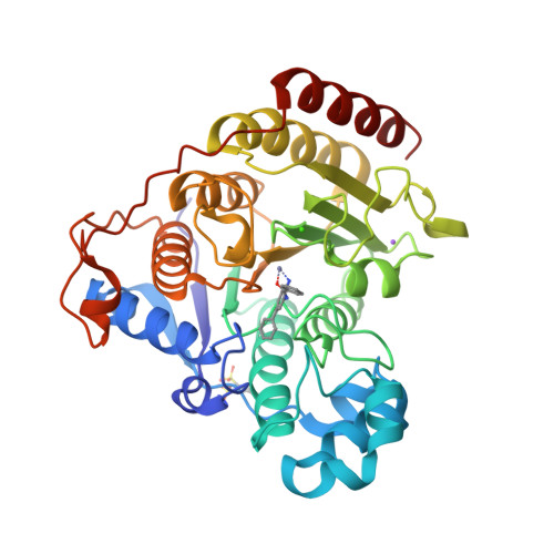
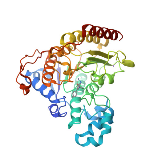
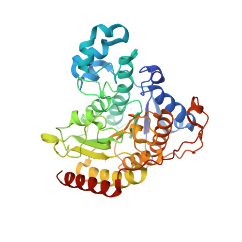
    A series of N-(2-amino-5-substituted phenyl)benzamides (3-21) were designed, synthesized and evaluated for their inhibition of HDAC2 and their cytotoxicity in HCT116 cancer cells. Multiple compounds from this series demonstrated time-dependent binding kinetics that is rationalized using a co-complex crystal structure of HDAC2 and N-(4-aminobiphenyl-3-yl)benzamide (6).


  • Organizational Affiliation

    Takeda San Diego, San Diego, CA 92121, USA.


Macromolecules
Find similar proteins by:  (by identity cutoff)  |  3D Structure
Entity ID: 1
MoleculeChains Sequence LengthOrganismDetailsImage
Histone deacetylase 2
A, B, C
367Homo sapiensMutation(s): 0 
Gene Names: HDAC2
EC: 3.5.1.98
UniProt & NIH Common Fund Data Resources
Find proteins for Q92769 (Homo sapiens)
Explore Q92769 
Go to UniProtKB:  Q92769
PHAROS:  Q92769
GTEx:  ENSG00000196591 
Entity Groups  
Sequence Clusters30% Identity50% Identity70% Identity90% Identity95% Identity100% Identity
UniProt GroupQ92769
Sequence Annotations
Expand
  • Reference Sequence
Small Molecules
Ligands 5 Unique
IDChains Name / Formula / InChI Key2D Diagram3D Interactions
LLX
Query on LLX

Download Ideal Coordinates CCD File 
G [auth A],
L [auth B],
P [auth C]
N-(4-aminobiphenyl-3-yl)benzamide
C19 H16 N2 O
ZWLFHHHQRUYIBT-UHFFFAOYSA-N
NHE
Query on NHE

Download Ideal Coordinates CCD File 
H [auth A]2-[N-CYCLOHEXYLAMINO]ETHANE SULFONIC ACID
C8 H17 N O3 S
MKWKNSIESPFAQN-UHFFFAOYSA-N
ZN
Query on ZN

Download Ideal Coordinates CCD File 
D [auth A],
I [auth B],
M [auth C]
ZINC ION
Zn
PTFCDOFLOPIGGS-UHFFFAOYSA-N
CA
Query on CA

Download Ideal Coordinates CCD File 
E [auth A],
J [auth B],
N [auth C]
CALCIUM ION
Ca
BHPQYMZQTOCNFJ-UHFFFAOYSA-N
NA
Query on NA

Download Ideal Coordinates CCD File 
F [auth A],
K [auth B],
O [auth C]
SODIUM ION
Na
FKNQFGJONOIPTF-UHFFFAOYSA-N
Binding Affinity Annotations 
IDSourceBinding Affinity
LLX PDBBind:  3MAX IC50: 900 (nM) from 1 assay(s)
BindingDB:  3MAX IC50: min: 27, max: 1200 (nM) from 9 assay(s)
Binding MOAD:  3MAX IC50: 27 (nM) from 1 assay(s)
Experimental Data & Validation

Experimental Data

  • Method: X-RAY DIFFRACTION
  • Resolution: 2.05 Å
  • R-Value Free: 0.204 
  • R-Value Work: 0.164 
  • R-Value Observed: 0.166 
  • Space Group: P 21 21 21
Unit Cell:
Length ( Å )Angle ( ˚ )
a = 92.311α = 90
b = 97.227β = 90
c = 138.849γ = 90
Software Package:
Software NamePurpose
HKL-2000data collection
MOLREPphasing
REFMACrefinement
HKL-2000data reduction
HKL-2000data scaling

Structure Validation

View Full Validation Report


Ligand Structure Quality Assessment 


Entry History 

Deposition Data

Revision History  (Full details and data files)

  • Version 1.0: 2010-04-28
    Type: Initial release
  • Version 1.1: 2011-07-13
    Changes: Version format compliance
  • Version 1.2: 2024-02-21
    Changes: Data collection, Database references, Derived calculations