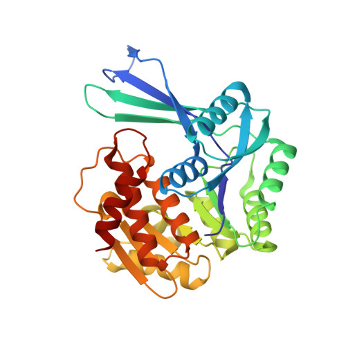
3N1C

Crystal structure of the phosphofructokinase-2 from Escherichia coli in complex with fructose-6-phosphate


Experimental Data Snapshot

  • Method: X-RAY DIFFRACTION
  • Resolution: 2.00 Å
  • R-Value Free: 0.221 
  • R-Value Work: 0.180 
  • R-Value Observed: 0.182 

wwPDB Validation   3D Report Full Report


Ligand Structure Quality Assessment 


This is version 1.3 of the entry. See complete history


Literature

The Crystal Complex of Phosphofructokinase-2 of Escherichia coli with Fructose-6-phosphate: KINETIC AND STRUCTURAL ANALYSIS OF THE ALLOSTERIC ATP INHIBITION.

Cabrera, R.Baez, M.Pereira, H.M.Caniuguir, A.Garratt, R.C.Babul, J.

(2011) J Biol Chem 286: 5774-5783

  • DOI: https://doi.org/10.1074/jbc.M110.163162
  • Primary Citation of Related Structures:  
    3N1C

  • PubMed Abstract: 

    Substrate inhibition by ATP is a regulatory feature of the phosphofructokinases isoenzymes from Escherichia coli (Pfk-1 and Pfk-2). Under gluconeogenic conditions, the loss of this regulation in Pfk-2 causes substrate cycling of fructose-6-phosphate (fructose-6-P) and futile consumption of ATP delaying growth. In the present work, we have broached the mechanism of ATP-induced inhibition of Pfk-2 from both structural and kinetic perspectives. The crystal structure of Pfk-2 in complex with fructose-6-P is reported to a resolution of 2 Å. The comparison of this structure with the previously reported inhibited form of the enzyme suggests a negative interplay between fructose-6-P binding and allosteric binding of MgATP. Initial velocity experiments show a linear increase of the apparent K(0.5) for fructose-6-P and a decrease in the apparent k(cat) as a function of MgATP concentration. These effects occur simultaneously with the induction of a sigmoidal kinetic behavior (n(H) of approximately 2). Differences and resemblances in the patterns of fructose-6-P binding and the mechanism of inhibition are discussed for Pfk-1 and Pfk-2, as an example of evolutionary convergence, because these enzymes do not share a common ancestor.


  • Organizational Affiliation

    Departamento de Biología, Facultad de Ciencias, Universidad de Chile, Santiago, Chile. ricabrer@uchile.cl


Macromolecules
Find similar proteins by:  (by identity cutoff)  |  3D Structure
Entity ID: 1
MoleculeChains Sequence LengthOrganismDetailsImage
6-phosphofructokinase isozyme 2
A, B, C, D
309Escherichia coli K-12Mutation(s): 0 
Gene Names: b1723JW5280PFK2pfkB
EC: 2.7.1.11
UniProt
Find proteins for P06999 (Escherichia coli (strain K12))
Explore P06999 
Go to UniProtKB:  P06999
Entity Groups  
Sequence Clusters30% Identity50% Identity70% Identity90% Identity95% Identity100% Identity
UniProt GroupP06999
Sequence Annotations
Expand
  • Reference Sequence
Experimental Data & Validation

Experimental Data

  • Method: X-RAY DIFFRACTION
  • Resolution: 2.00 Å
  • R-Value Free: 0.221 
  • R-Value Work: 0.180 
  • R-Value Observed: 0.182 
  • Space Group: C 2 2 21
Unit Cell:
Length ( Å )Angle ( ˚ )
a = 68.793α = 90
b = 153.605β = 90
c = 223.79γ = 90
Software Package:
Software NamePurpose
MOSFLMdata reduction
SCALAdata scaling
PHASERphasing
PHENIXrefinement
PDB_EXTRACTdata extraction

Structure Validation

View Full Validation Report


Ligand Structure Quality Assessment 


Entry History 

Deposition Data

Revision History  (Full details and data files)

  • Version 1.0: 2010-12-08
    Type: Initial release
  • Version 1.1: 2011-07-13
    Changes: Version format compliance
  • Version 1.2: 2020-07-29
    Type: Remediation
    Reason: Carbohydrate remediation
    Changes: Advisory, Data collection, Derived calculations, Structure summary
  • Version 1.3: 2023-09-06
    Changes: Data collection, Database references, Refinement description, Structure summary