3O62

Nucleosome core particle modified with a cisplatin 1,3-cis-{Pt(NH3)2}2+-d(GpTpG) intrastrand cross-link


Experimental Data Snapshot

  • Method: X-RAY DIFFRACTION
  • Resolution: 3.22 Å
  • R-Value Free: 0.306 
  • R-Value Work: 0.249 
  • R-Value Observed: 0.252 

wwPDB Validation   3D Report Full Report


This is version 1.2 of the entry. See complete history


Literature

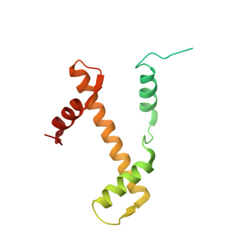
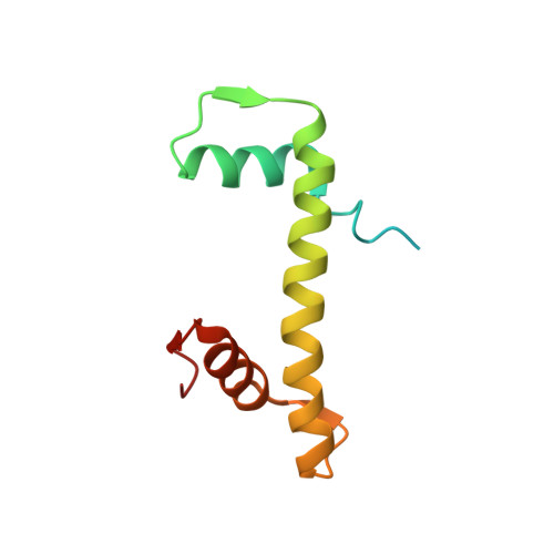
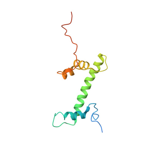
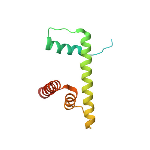
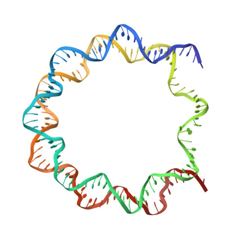
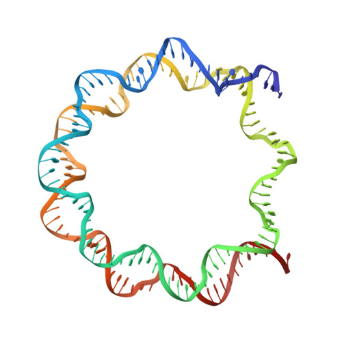
Consequences of Cisplatin binding on nucleosome structure and dynamics.

Todd, R.C.Lippard, S.J.

(2010) Chem Biol 17: 1334-1343

  • DOI: https://doi.org/10.1016/j.chembiol.2010.10.018
  • Primary Citation of Related Structures:  
    3O62

  • PubMed Abstract: 

    The effects of cisplatin binding to DNA were explored at the nucleosome level to incorporate key features of the eukaryotic nuclear environment. An X-ray crystal structure of a site-specifically platinated nucleosome carrying a 1,3-cis-{Pt(NH₃)₂}²+-d(GpTpG) intrastrand cross-link reveals the details of how this adduct dictates the rotational positioning of DNA in the nucleosome. Results from in vitro nucleosome mobility assays indicate that a single platinum adduct interferes with ATP-independent sliding of DNA around the octamer core. Data from in vitro transcription experiments suggest that RNA polymerases can successfully navigate along cisplatin-damaged DNA templates that contain nucleosomes, but stall when the transcription elongation complex physically contacts a platinum cross-link located on the template strand. These results provide information about the effects of cisplatin binding to nuclear DNA and enhance our understanding of the mechanism of transcription inhibition by platinum anticancer compounds.


  • Organizational Affiliation

    Department of Chemistry, Massachusetts Institute of Technology, Cambridge, MA 02139, USA.


Macromolecules

Find similar proteins by:  (by identity cutoff)  |  3D Structure
Entity ID: 1
MoleculeChains Sequence LengthOrganismDetailsImage
Histone H3.2
A, E
135Xenopus laevisMutation(s): 0 
Gene Names: h3.2
UniProt
Find proteins for P84233 (Xenopus laevis)
Explore P84233 
Go to UniProtKB:  P84233
Entity Groups  
Sequence Clusters30% Identity50% Identity70% Identity90% Identity95% Identity100% Identity
UniProt GroupP84233
Sequence Annotations
Expand
  • Reference Sequence
Find similar proteins by:  (by identity cutoff)  |  3D Structure
Entity ID: 2
MoleculeChains Sequence LengthOrganismDetailsImage
Histone H4
B, F
102Xenopus laevisMutation(s): 0 
UniProt
Find proteins for P62799 (Xenopus laevis)
Explore P62799 
Go to UniProtKB:  P62799
Entity Groups  
Sequence Clusters30% Identity50% Identity70% Identity90% Identity95% Identity100% Identity
UniProt GroupP62799
Sequence Annotations
Expand
  • Reference Sequence
Find similar proteins by:  (by identity cutoff)  |  3D Structure
Entity ID: 3
MoleculeChains Sequence LengthOrganismDetailsImage
Histone H2A type 1
C, G
128Xenopus laevisMutation(s): 0 
UniProt
Find proteins for P06897 (Xenopus laevis)
Explore P06897 
Go to UniProtKB:  P06897
Entity Groups  
Sequence Clusters30% Identity50% Identity70% Identity90% Identity95% Identity100% Identity
UniProt GroupP06897
Sequence Annotations
Expand
  • Reference Sequence
Find similar proteins by:  (by identity cutoff)  |  3D Structure
Entity ID: 4
MoleculeChains Sequence LengthOrganismDetailsImage
Histone H2B 1.1
D, H
122Xenopus laevisMutation(s): 0 
UniProt
Find proteins for P02281 (Xenopus laevis)
Explore P02281 
Go to UniProtKB:  P02281
Entity Groups  
Sequence Clusters30% Identity50% Identity70% Identity90% Identity95% Identity100% Identity
UniProt GroupP02281
Sequence Annotations
Expand
  • Reference Sequence
Find similar nucleic acids by:  (by identity cutoff)  |  3D Structure
Entity ID: 5
MoleculeChains LengthOrganismImage
DNA (146-MER)146N/A
Sequence Annotations
Expand
  • Reference Sequence
Find similar nucleic acids by:  (by identity cutoff)  |  3D Structure
Entity ID: 6
MoleculeChains LengthOrganismImage
DNA (146-MER)146N/A
Sequence Annotations
Expand
  • Reference Sequence
Small Molecules
Ligands 1 Unique
IDChains Name / Formula / InChI Key2D Diagram3D Interactions
CPT
Query on CPT

Download Ideal Coordinates CCD File 
K [auth I]Cisplatin
Cl2 H6 N2 Pt
LXZZYRPGZAFOLE-UHFFFAOYSA-L
Experimental Data & Validation

Experimental Data

  • Method: X-RAY DIFFRACTION
  • Resolution: 3.22 Å
  • R-Value Free: 0.306 
  • R-Value Work: 0.249 
  • R-Value Observed: 0.252 
  • Space Group: P 21 21 21
Unit Cell:
Length ( Å )Angle ( ˚ )
a = 106.095α = 90
b = 109.551β = 90
c = 177.176γ = 90
Software Package:
Software NamePurpose
HKL-2000data collection
PHASERphasing
REFMACrefinement
HKL-2000data reduction
HKL-2000data scaling

Structure Validation

View Full Validation Report


Entry History 

Deposition Data

Revision History  (Full details and data files)

  • Version 1.0: 2011-01-05
    Type: Initial release
  • Version 1.1: 2011-07-13
    Changes: Version format compliance
  • Version 1.2: 2024-02-21
    Changes: Data collection, Database references, Derived calculations