3OZU

The Crystal Structure of flavohemoglobin from R. eutrophus in complex with miconazole


Experimental Data Snapshot

  • Method: X-RAY DIFFRACTION
  • Resolution: 2.00 Å
  • R-Value Free: 0.233 
  • R-Value Work: 0.185 
  • R-Value Observed: 0.188 

wwPDB Validation   3D Report Full Report


Ligand Structure Quality Assessment 


This is version 1.2 of the entry. See complete history


Literature

Structure of Ralstonia eutropha Flavohemoglobin in Complex with Three Antibiotic Azole Compounds.

El Hammi, E.Warkentin, E.Demmer, U.Limam, F.Marzouki, N.M.Ermler, U.Baciou, L.

(2011) Biochemistry 50: 1255-1264

  • DOI: https://doi.org/10.1021/bi101650q
  • Primary Citation of Related Structures:  
    3OZU, 3OZV, 3OZW

  • PubMed Abstract: 

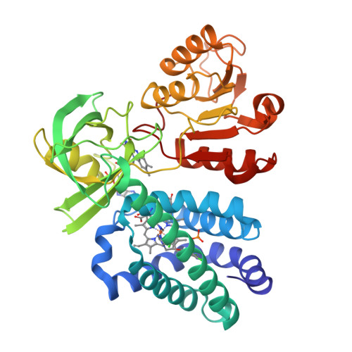
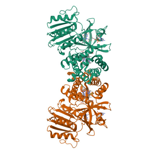
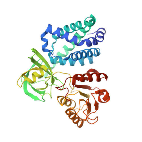
    Flavohemoglobins (flavoHbs) are enzymes that operate primarily as nitric oxide dioxygenases and shuttle thereby electrons among NAD(P)H, FAD, heme, and a ligated redox-active substrate such as O(2). They function in the bacterial defense against nitrosative stress and are therefore considered as targets for new antibiotic drugs. Recently, azole derivatives were proven to be attractive nitric oxide dioxygenase inhibitors, and to explore their binding characteristics, we determined the X-ray structure of the flavoHb from Ralstonia eutropha in a complex with miconazole (FHP(M)), econazole (FHP(E)), and ketoconazole (FHP(K)). In agreement with UV-vis spectroscopic data, one azole compound binds inside the distal heme pocket and ligates to the heme iron by its imidazole substituent. The two additional substituents, mostly chlorinated phenyl groups, form a series of van der Waals contacts with the protein matrix. Both interactions explain their high affinity for flavoHbs, the binding constants being 2.6, 1.2, and 11.6 μM for miconazole, econazole, and ketoconazole, respectively. The FHP(M) and FHP(Lip) (flavoHbs originally loaded with a phospholipid) structures share an "open" state and the FHP(E) and FHP(K) structures a "closed" state. Although the azole compounds were able to push the lipid out of its binding site, a fatty acid fragment is still bound inside the heme pocket of FHP(E) and FHP(K) and dictates the state of the protein. The ligand-induced open-to-closed transition involves a reorientation of the NADH domain accompanied by conformational changes in the C-terminal arm, helix E, and the CE loop resulting in an encapsulation of the heme-binding pocket. Implications of the observed open-to-closed process on the catalytic cycle are discussed.


  • Organizational Affiliation

    Laboratoire de Chimie Physique, CNRS-Université Paris-Sud 11, UMR8000, F-91405 Orsay, France.


Macromolecules
Find similar proteins by:  (by identity cutoff)  |  3D Structure
Entity ID: 1
MoleculeChains Sequence LengthOrganismDetailsImage
Flavohemoprotein403Cupriavidus necator H16Mutation(s): 0 
Gene Names: hmpfhpPHG200
EC: 1.14.12.17
UniProt
Find proteins for P39662 (Cupriavidus necator (strain ATCC 17699 / DSM 428 / KCTC 22496 / NCIMB 10442 / H16 / Stanier 337))
Explore P39662 
Go to UniProtKB:  P39662
Entity Groups  
Sequence Clusters30% Identity50% Identity70% Identity90% Identity95% Identity100% Identity
UniProt GroupP39662
Sequence Annotations
Expand
  • Reference Sequence
Small Molecules
Ligands 4 Unique
IDChains Name / Formula / InChI Key2D Diagram3D Interactions
FAD
Query on FAD

Download Ideal Coordinates CCD File 
C [auth A]FLAVIN-ADENINE DINUCLEOTIDE
C27 H33 N9 O15 P2
VWWQXMAJTJZDQX-UYBVJOGSSA-N
HEM
Query on HEM

Download Ideal Coordinates CCD File 
B [auth A]PROTOPORPHYRIN IX CONTAINING FE
C34 H32 Fe N4 O4
KABFMIBPWCXCRK-RGGAHWMASA-L
X89
Query on X89

Download Ideal Coordinates CCD File 
D [auth A]1-[(2R)-2-[(2,4-dichlorobenzyl)oxy]-2-(2,4-dichlorophenyl)ethyl]-1H-imidazole
C18 H14 Cl4 N2 O
BYBLEWFAAKGYCD-SFHVURJKSA-N
PO4
Query on PO4

Download Ideal Coordinates CCD File 
E [auth A]PHOSPHATE ION
O4 P
NBIIXXVUZAFLBC-UHFFFAOYSA-K
Binding Affinity Annotations 
IDSourceBinding Affinity
X89 Binding MOAD:  3OZU Kd: 2600 (nM) from 1 assay(s)
Experimental Data & Validation

Experimental Data

  • Method: X-RAY DIFFRACTION
  • Resolution: 2.00 Å
  • R-Value Free: 0.233 
  • R-Value Work: 0.185 
  • R-Value Observed: 0.188 
  • Space Group: C 1 2 1
Unit Cell:
Length ( Å )Angle ( ˚ )
a = 121.339α = 90
b = 105.278β = 101.9
c = 46.445γ = 90
Software Package:
Software NamePurpose
MAR345dtbdata collection
EPMRphasing
REFMACrefinement
XDSdata reduction
XDSdata scaling

Structure Validation

View Full Validation Report


Ligand Structure Quality Assessment 


Entry History 

Deposition Data

Revision History  (Full details and data files)

  • Version 1.0: 2011-03-16
    Type: Initial release
  • Version 1.1: 2011-07-13
    Changes: Version format compliance
  • Version 1.2: 2023-09-06
    Changes: Data collection, Database references, Derived calculations, Refinement description