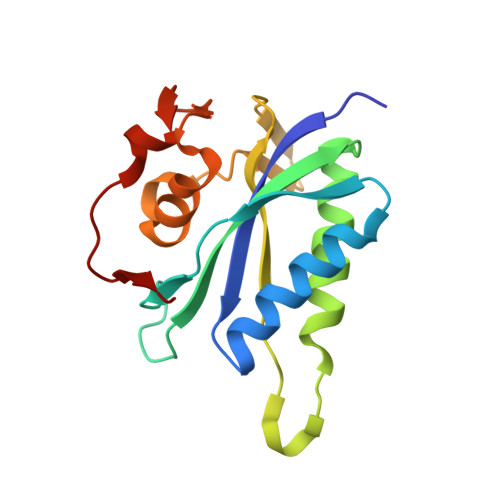
Structure of S. aureus HPPK and the discovery of a new substrate site inhibitor
Chhabra, S., Dolezal, O., Collins, B.M., Newman, J., Simpson, J.S., Macreadie, I.G., Fernley, R., Peat, T.S., Swarbrick, J.D.(2012) PLoS One 7: e29444-e29444
- PubMed: 22276115
- DOI: https://doi.org/10.1371/journal.pone.0029444
- Primary Citation of Related Structures:
3QBC - PubMed Abstract:
The first structural and biophysical data on the folate biosynthesis pathway enzyme and drug target, 6-hydroxymethyl-7,8-dihydropterin pyrophosphokinase (SaHPPK), from the pathogen Staphylococcus aureus is presented. HPPK is the second essential enzyme in the pathway catalysing the pyrophosphoryl transfer from cofactor (ATP) to the substrate (6-hydroxymethyl-7,8-dihydropterin, HMDP). In-silico screening identified 8-mercaptoguanine which was shown to bind with an equilibrium dissociation constant, K(d), of ∼13 µM as measured by isothermal titration calorimetry (ITC) and surface plasmon resonance (SPR). An IC(50) of ∼41 µM was determined by means of a luminescent kinase assay. In contrast to the biological substrate, the inhibitor has no requirement for magnesium or the ATP cofactor for competitive binding to the substrate site. The 1.65 Å resolution crystal structure of the inhibited complex showed that it binds in the pterin site and shares many of the key intermolecular interactions of the substrate. Chemical shift and (15)N heteronuclear NMR measurements reveal that the fast motion of the pterin-binding loop (L2) is partially dampened in the SaHPPK/HMDP/α,β-methylene adenosine 5'-triphosphate (AMPCPP) ternary complex, but the ATP loop (L3) remains mobile on the µs-ms timescale. In contrast, for the SaHPPK/8-mercaptoguanine/AMPCPP ternary complex, the loop L2 becomes rigid on the fast timescale and the L3 loop also becomes more ordered--an observation that correlates with the large entropic penalty associated with inhibitor binding as revealed by ITC. NMR data, including (15)N-(1)H residual dipolar coupling measurements, indicate that the sulfur atom in the inhibitor is important for stabilizing and restricting important motions of the L2 and L3 catalytic loops in the inhibited ternary complex. This work describes a comprehensive analysis of a new HPPK inhibitor, and may provide a foundation for the development of novel antimicrobials targeting the folate biosynthetic pathway.
Organizational Affiliation:
Medicinal Chemistry and Drug Action, Monash Institute of Pharmaceutical Sciences, Monash University, Parkville, Australia.