3RGW

Crystal structure at 1.5 A resolution of an H2-reduced, O2-tolerant hydrogenase from Ralstonia eutropha unmasks a novel iron-sulfur cluster


Experimental Data Snapshot

  • Method: X-RAY DIFFRACTION
  • Resolution: 1.50 Å
  • R-Value Free: 0.153 
  • R-Value Work: 0.140 
  • R-Value Observed: 0.140 

wwPDB Validation   3D Report Full Report


This is version 1.3 of the entry. See complete history


Literature

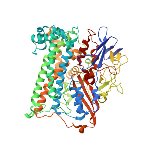
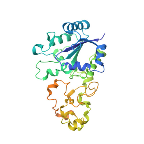
The crystal structure of an oxygen-tolerant hydrogenase uncovers a novel iron-sulphur centre.

Fritsch, J.Scheerer, P.Frielingsdorf, S.Kroschinsky, S.Friedrich, B.Lenz, O.Spahn, C.M.

(2011) Nature 479: 249-252

  • DOI: https://doi.org/10.1038/nature10505
  • Primary Citation of Related Structures:  
    3RGW

  • PubMed Abstract: 

    Hydrogenases are abundant enzymes that catalyse the reversible interconversion of H(2) into protons and electrons at high rates. Those hydrogenases maintaining their activity in the presence of O(2) are considered to be central to H(2)-based technologies, such as enzymatic fuel cells and for light-driven H(2) production. Despite comprehensive genetic, biochemical, electrochemical and spectroscopic investigations, the molecular background allowing a structural interpretation of how the catalytic centre is protected from irreversible inactivation by O(2) has remained unclear. Here we present the crystal structure of an O(2)-tolerant [NiFe]-hydrogenase from the aerobic H(2) oxidizer Ralstonia eutropha H16 at 1.5 Å resolution. The heterodimeric enzyme consists of a large subunit harbouring the catalytic centre in the H(2)-reduced state and a small subunit containing an electron relay consisting of three different iron-sulphur clusters. The cluster proximal to the active site displays an unprecedented [4Fe-3S] structure and is coordinated by six cysteines. According to the current model, this cofactor operates as an electronic switch depending on the nature of the gas molecule approaching the active site. It serves as an electron acceptor in the course of H(2) oxidation and as an electron-delivering device upon O(2) attack at the active site. This dual function is supported by the capability of the novel iron-sulphur cluster to adopt three redox states at physiological redox potentials. The second structural feature is a network of extended water cavities that may act as a channel facilitating the removal of water produced at the [NiFe] active site. These discoveries will have an impact on the design of biological and chemical H(2)-converting catalysts that are capable of cycling H(2) in air.


  • Organizational Affiliation

    Mikrobiologie, Institut für Biologie, Humboldt-Universität zu Berlin, Chausseestraße 117, 10115 Berlin, Germany.


Macromolecules
Find similar proteins by:  (by identity cutoff)  |  3D Structure
Entity ID: 1
MoleculeChains Sequence LengthOrganismDetailsImage
Membrane-bound hydrogenase (NIFE) large subunit HOXGA [auth L]603Cupriavidus necator H16Mutation(s): 0 
EC: 1.12.99.6
UniProt
Find proteins for P31891 (Cupriavidus necator (strain ATCC 17699 / DSM 428 / KCTC 22496 / NCIMB 10442 / H16 / Stanier 337))
Explore P31891 
Go to UniProtKB:  P31891
Entity Groups  
Sequence Clusters30% Identity50% Identity70% Identity90% Identity95% Identity100% Identity
UniProt GroupP31891
Sequence Annotations
Expand
  • Reference Sequence
Find similar proteins by:  (by identity cutoff)  |  3D Structure
Entity ID: 2
MoleculeChains Sequence LengthOrganismDetailsImage
Membrane-bound hydrogenase (NIFE) small subunit HOXKB [auth S]339Cupriavidus necator H16Mutation(s): 0 
EC: 1.12.99.6
UniProt
Find proteins for P31892 (Cupriavidus necator (strain ATCC 17699 / DSM 428 / KCTC 22496 / NCIMB 10442 / H16 / Stanier 337))
Explore P31892 
Go to UniProtKB:  P31892
Entity Groups  
Sequence Clusters30% Identity50% Identity70% Identity90% Identity95% Identity100% Identity
UniProt GroupP31892
Sequence Annotations
Expand
  • Reference Sequence
Small Molecules
Ligands 5 Unique
IDChains Name / Formula / InChI Key2D Diagram3D Interactions
SF4
Query on SF4

Download Ideal Coordinates CCD File 
E [auth S]IRON/SULFUR CLUSTER
Fe4 S4
LJBDFODJNLIPKO-UHFFFAOYSA-N
F4S
Query on F4S

Download Ideal Coordinates CCD File 
G [auth S]FE4-S3 CLUSTER
Fe4 S3
QQACTBFBZNWJMV-UHFFFAOYSA-N
F3S
Query on F3S

Download Ideal Coordinates CCD File 
F [auth S]FE3-S4 CLUSTER
Fe3 S4
FCXHZBQOKRZXKS-UHFFFAOYSA-N
NFU
Query on NFU

Download Ideal Coordinates CCD File 
C [auth L]formyl[bis(hydrocyanato-1kappaC)]ironnickel(Fe-Ni)
C3 H Fe N2 Ni O
QCZROEOIPZWDEO-UHFFFAOYSA-N
MG
Query on MG

Download Ideal Coordinates CCD File 
D [auth L]MAGNESIUM ION
Mg
JLVVSXFLKOJNIY-UHFFFAOYSA-N
Experimental Data & Validation

Experimental Data

  • Method: X-RAY DIFFRACTION
  • Resolution: 1.50 Å
  • R-Value Free: 0.153 
  • R-Value Work: 0.140 
  • R-Value Observed: 0.140 
  • Space Group: P 21 21 21
Unit Cell:
Length ( Å )Angle ( ˚ )
a = 73.091α = 90
b = 95.646β = 90
c = 119.145γ = 90
Software Package:
Software NamePurpose
MxCuBEdata collection
PHASERphasing
REFMACrefinement
XDSdata reduction
SCALAdata scaling

Structure Validation

View Full Validation Report


Entry History 

Deposition Data

Revision History  (Full details and data files)

  • Version 1.0: 2011-10-26
    Type: Initial release
  • Version 1.1: 2011-11-16
    Changes: Database references
  • Version 1.2: 2011-11-23
    Changes: Database references
  • Version 1.3: 2023-09-13
    Changes: Data collection, Database references, Derived calculations, Refinement description