3US0

Structure of p63 DNA Binding Domain in Complex with a 22 Base Pair A/T Rich Response Element Containing a Two Base Pair "AT" Spacer Between Half Sites


Experimental Data Snapshot

  • Method: X-RAY DIFFRACTION
  • Resolution: 2.50 Å
  • R-Value Free: 0.239 
  • R-Value Work: 0.206 
  • R-Value Observed: 0.208 

wwPDB Validation   3D Report Full Report


This is version 1.3 of the entry. See complete history


Literature

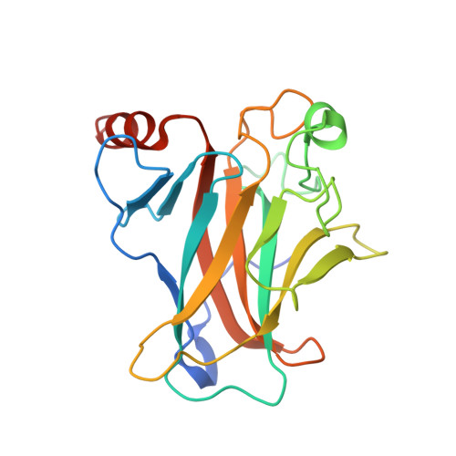
Pliable DNA Conformation of Response Elements Bound to Transcription Factor p63.

Chen, C.Gorlatova, N.Herzberg, O.

(2012) J Biol Chem 287: 7477-7486

  • DOI: https://doi.org/10.1074/jbc.M111.315820
  • Primary Citation of Related Structures:  
    3US0, 3US1, 3US2

  • PubMed Abstract: 

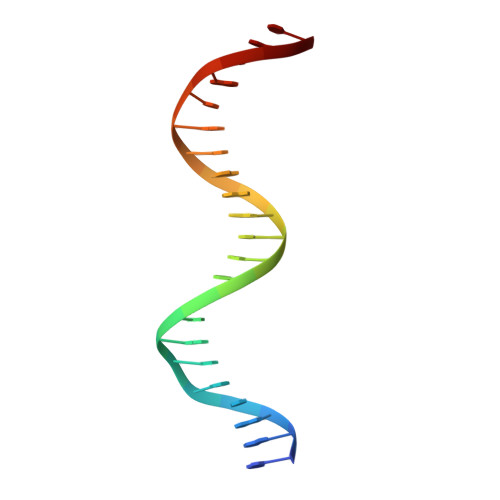
    We show that changes in the nucleotide sequence alter the DNA conformation in the crystal structures of p63 DNA-binding domain (p63DBD) bound to its response element. The conformation of a 22-bp canonical response element containing an AT spacer between the two half-sites is unaltered compared with that containing a TA spacer, exhibiting superhelical trajectory. In contrast, a GC spacers abolishes the DNA superhelical trajectory and exhibits less bent DNA, suggesting that increased GC content accompanies increased double helix rigidity. A 19-bp DNA, representing an AT-rich response element with overlapping half-sites, maintains superhelical trajectory and reveals two interacting p63DBD dimers crossing one another at 120°. p63DBD binding assays to response elements of increasing length complement the structural studies. We propose that DNA deformation may affect promoter activity, that the ability of p63DBD to bind to superhelical DNA suggests that it is capable of binding to nucleosomes, and that overlapping response elements may provide a mechanism to distinguish between p63 and p53 promoters.


  • Organizational Affiliation

    W. M. Keck Laboratory for Structural Biology, Institute for Bioscience and Biotechnology Research and the Department of Chemistry and Biochemistry, University of Maryland, Rockville, Maryland 20850, USA.


Macromolecules

Find similar proteins by:  (by identity cutoff)  |  3D Structure
Entity ID: 1
MoleculeChains Sequence LengthOrganismDetailsImage
Tumor protein 63
A, B, C, D
203Homo sapiensMutation(s): 0 
Gene Names: KETP63P73HP73LTP63TP73L
UniProt & NIH Common Fund Data Resources
Find proteins for Q9H3D4 (Homo sapiens)
Explore Q9H3D4 
Go to UniProtKB:  Q9H3D4
PHAROS:  Q9H3D4
GTEx:  ENSG00000073282 
Entity Groups  
Sequence Clusters30% Identity50% Identity70% Identity90% Identity95% Identity100% Identity
UniProt GroupQ9H3D4
Sequence Annotations
Expand
  • Reference Sequence

Find similar nucleic acids by:  Sequence   |   3D Structure  

Entity ID: 2
MoleculeChains LengthOrganismImage
5'-D(*AP*AP*AP*CP*AP*TP*GP*TP*TP*TP*AP*TP*AP*AP*AP*CP*AP*TP*GP*TP*TP*T)-3'
E, F
22N/A
Sequence Annotations
Expand
  • Reference Sequence
Experimental Data & Validation

Experimental Data

  • Method: X-RAY DIFFRACTION
  • Resolution: 2.50 Å
  • R-Value Free: 0.239 
  • R-Value Work: 0.206 
  • R-Value Observed: 0.208 
  • Space Group: P 63
Unit Cell:
Length ( Å )Angle ( ˚ )
a = 141.806α = 90
b = 141.806β = 90
c = 119.604γ = 120
Software Package:
Software NamePurpose
JBluIce-EPICSdata collection
PHASERphasing
PHENIXrefinement
XDSdata reduction
XSCALEdata scaling

Structure Validation

View Full Validation Report


Entry History 

Deposition Data

Revision History  (Full details and data files)

  • Version 1.0: 2012-02-01
    Type: Initial release
  • Version 1.1: 2012-03-21
    Changes: Database references
  • Version 1.2: 2017-11-08
    Changes: Refinement description
  • Version 1.3: 2023-09-13
    Changes: Data collection, Database references, Derived calculations, Refinement description