3UT5

Tubulin-Colchicine-Ustiloxin: Stathmin-like domain complex


Experimental Data Snapshot

  • Method: X-RAY DIFFRACTION
  • Resolution: 2.73 Å
  • R-Value Free: 0.222 
  • R-Value Work: 0.184 
  • R-Value Observed: 0.186 

wwPDB Validation   3D Report Full Report


Ligand Structure Quality Assessment 


This is version 2.0 of the entry. See complete history


Literature

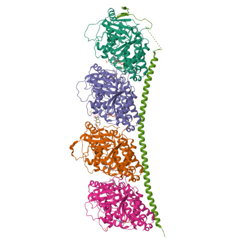
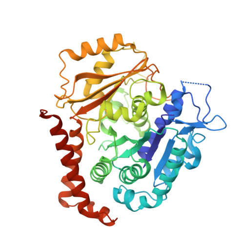
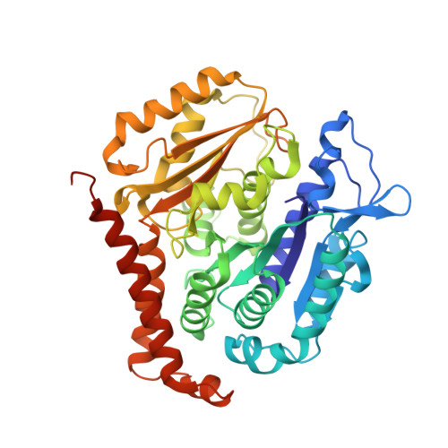
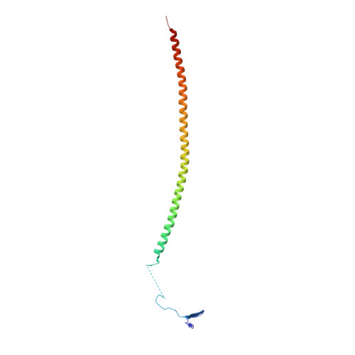
Structural plasticity of tubulin assembly probed by vinca-domain ligands.

Ranaivoson, F.M.Gigant, B.Berritt, S.Joullie, M.Knossow, M.

(2012) Acta Crystallogr D Biol Crystallogr 68: 927-934

  • DOI: https://doi.org/10.1107/S0907444912017143
  • Primary Citation of Related Structures:  
    3UT5, 4EB6

  • PubMed Abstract: 

    Vinca-domain ligands are compounds that bind to tubulin at its inter-heterodimeric interface and favour heterogeneous protofilament-like assemblies, giving rise to helices and rings. This is the basis for their inhibition of microtubule assembly, for their antimitotic activities and for their use in anticancer chemotherapy. Ustiloxins are vinca-domain ligands with a well established total synthesis. A 2.7 Å resolution structure of ustiloxin D bound to the vinca domain embedded in the complex of two tubulins with the stathmin-like domain of RB3 (T(2)R) has been determined. This finding precisely defines the interactions of ustiloxins with tubulin and, taken together with structures of other vinca-ligand complexes, allows structure-based suggestions to be made for improved activity. These comparisons also provide a rationale for the large-scale polymorphism of the protofilament-like assemblies mediated by vinca-domain ligands based on local differences in their interactions with the two tubulin heterodimers constituting their binding site.


  • Organizational Affiliation

    Laboratoire d'Enzymologie et Biochimie Structurales, Centre de Recherche de Gif, CNRS, 91198 Gif-sur-Yvette, France.


Macromolecules
Find similar proteins by:  (by identity cutoff)  |  3D Structure
Entity ID: 1
MoleculeChains Sequence LengthOrganismDetailsImage
Tubulin alpha chain
A, C
451Ovis ariesMutation(s): 0 
UniProt
Find proteins for D0VWZ0 (Ovis aries)
Explore D0VWZ0 
Go to UniProtKB:  D0VWZ0
Entity Groups  
Sequence Clusters30% Identity50% Identity70% Identity90% Identity95% Identity100% Identity
UniProt GroupD0VWZ0
Sequence Annotations
Expand
  • Reference Sequence
Find similar proteins by:  (by identity cutoff)  |  3D Structure
Entity ID: 2
MoleculeChains Sequence LengthOrganismDetailsImage
Tubulin beta chain
B, D
445Ovis ariesMutation(s): 0 
UniProt
Find proteins for D0VWY9 (Ovis aries)
Explore D0VWY9 
Go to UniProtKB:  D0VWY9
Entity Groups  
Sequence Clusters30% Identity50% Identity70% Identity90% Identity95% Identity100% Identity
UniProt GroupD0VWY9
Sequence Annotations
Expand
  • Reference Sequence
Find similar proteins by:  (by identity cutoff)  |  3D Structure
Entity ID: 3
MoleculeChains Sequence LengthOrganismDetailsImage
Stathmin-4142Rattus norvegicusMutation(s): 2 
Gene Names: Stmn4
UniProt
Find proteins for P63043 (Rattus norvegicus)
Explore P63043 
Go to UniProtKB:  P63043
Entity Groups  
Sequence Clusters30% Identity50% Identity70% Identity90% Identity95% Identity100% Identity
UniProt GroupP63043
Sequence Annotations
Expand
  • Reference Sequence

Find similar proteins by:  Sequence   |   3D Structure  

Entity ID: 4
MoleculeChains Sequence LengthOrganismDetailsImage
Vinca tetrapeptide4N/AMutation(s): 0 
Sequence Annotations
Expand
  • Reference Sequence
Small Molecules
Ligands 5 Unique
IDChains Name / Formula / InChI Key2D Diagram3D Interactions
GTP
Query on GTP

Download Ideal Coordinates CCD File 
G [auth A],
N [auth C]
GUANOSINE-5'-TRIPHOSPHATE
C10 H16 N5 O14 P3
XKMLYUALXHKNFT-UUOKFMHZSA-N
GDP
Query on GDP

Download Ideal Coordinates CCD File 
K [auth B],
P [auth D]
GUANOSINE-5'-DIPHOSPHATE
C10 H15 N5 O11 P2
QGWNDRXFNXRZMB-UUOKFMHZSA-N
LOC
Query on LOC

Download Ideal Coordinates CCD File 
L [auth B],
Q [auth D]
N-[(7S)-1,2,3,10-tetramethoxy-9-oxo-6,7-dihydro-5H-benzo[d]heptalen-7-yl]ethanamide
C22 H25 N O6
IAKHMKGGTNLKSZ-INIZCTEOSA-N
SO4
Query on SO4

Download Ideal Coordinates CCD File 
I [auth A],
J [auth A],
M [auth B],
R [auth D],
S [auth D]
SULFATE ION
O4 S
QAOWNCQODCNURD-UHFFFAOYSA-L
MG
Query on MG

Download Ideal Coordinates CCD File 
H [auth A],
O [auth C]
MAGNESIUM ION
Mg
JLVVSXFLKOJNIY-UHFFFAOYSA-N
Modified Residues  2 Unique
IDChains TypeFormula2D DiagramParent
0E5
Query on 0E5
F
L-PEPTIDE LINKINGC6 H13 N O3THR
0EA
Query on 0EA
F
L-PEPTIDE LINKINGC10 H13 N O5TYR
Binding Affinity Annotations 
IDSourceBinding Affinity
LOC BindingDB:  3UT5 Ki: min: 1.6, max: 5750 (nM) from 4 assay(s)
Kd: min: 3700, max: 1.13e+4 (nM) from 2 assay(s)
IC50: min: 6.7, max: 2.93e+4 (nM) from 24 assay(s)
Experimental Data & Validation

Experimental Data

  • Method: X-RAY DIFFRACTION
  • Resolution: 2.73 Å
  • R-Value Free: 0.222 
  • R-Value Work: 0.184 
  • R-Value Observed: 0.186 
  • Space Group: P 21 21 21
Unit Cell:
Length ( Å )Angle ( ˚ )
a = 64.81α = 90
b = 128.87β = 90
c = 254.84γ = 90
Software Package:
Software NamePurpose
ADSCdata collection
AMoREphasing
BUSTERrefinement
XDSdata reduction
SCALAdata scaling

Structure Validation

View Full Validation Report


Ligand Structure Quality Assessment 


Entry History 

Deposition Data

Revision History  (Full details and data files)

  • Version 1.0: 2012-08-01
    Type: Initial release
  • Version 1.1: 2012-12-12
    Changes: Other
  • Version 1.2: 2013-01-23
    Changes: Database references
  • Version 2.0: 2023-11-15
    Changes: Atomic model, Data collection, Database references, Derived calculations