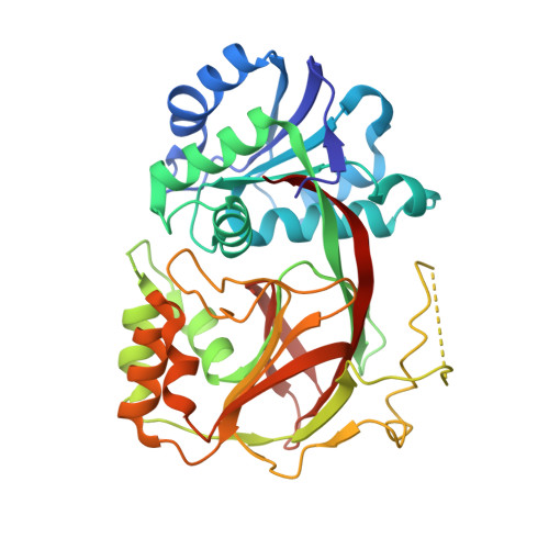
3VZC

Crystal structure of Sphingosine Kinase 1 with inhibitor


Experimental Data Snapshot

  • Method: X-RAY DIFFRACTION
  • Resolution: 2.30 Å
  • R-Value Free: 0.285 
  • R-Value Work: 0.229 
  • R-Value Observed: 0.232 

wwPDB Validation   3D Report Full Report


Ligand Structure Quality Assessment 


This is version 1.2 of the entry. See complete history


Literature

Molecular basis of sphingosine kinase 1 substrate recognition and catalysis.

Wang, Z.Min, X.Xiao, S.H.Johnstone, S.Romanow, W.Meininger, D.Xu, H.Liu, J.Dai, J.An, S.Thibault, S.Walker, N.

(2013) Structure 21: 798-809

  • DOI: https://doi.org/10.1016/j.str.2013.02.025
  • Primary Citation of Related Structures:  
    3VZB, 3VZC, 3VZD

  • PubMed Abstract: 

    Sphingosine kinase 1 (SphK1) is a lipid kinase that catalyzes the conversion of sphingosine to sphingosine-1-phosphate (S1P), which has been shown to play a role in lymphocyte trafficking, angiogenesis, and response to apoptotic stimuli. As a central enzyme in modulating the S1P levels in cells, SphK1 emerges as an important regulator for diverse cellular functions and a potential target for drug discovery. Here, we present the crystal structures of human SphK1 in the apo form and in complexes with a substrate sphingosine-like lipid, ADP, and an inhibitor at 2.0-2.3 Å resolution. The SphK1 structures reveal a two-domain architecture in which its catalytic site is located in the cleft between the two domains and a hydrophobic lipid-binding pocket is buried in the C-terminal domain. Comparative analysis of these structures with mutagenesis and kinetic studies provides insight into how SphK1 recognizes the lipid substrate and catalyzes ATP-dependent phosphorylation.


  • Organizational Affiliation

    Department of Molecular Structure and Characterization, Amgen, Inc., 1120 Veterans Boulevard, South San Francisco, CA 94080, USA. zwang@amgen.com


Macromolecules
Find similar proteins by:  (by identity cutoff)  |  3D Structure
Entity ID: 1
MoleculeChains Sequence LengthOrganismDetailsImage
Sphingosine kinase 1
A, B, C, D, E
A, B, C, D, E, F
361Homo sapiensMutation(s): 0 
Gene Names: SPHK1SPHKSPK
EC: 2.7.1.91
UniProt & NIH Common Fund Data Resources
Find proteins for Q9NYA1 (Homo sapiens)
Explore Q9NYA1 
Go to UniProtKB:  Q9NYA1
PHAROS:  Q9NYA1
GTEx:  ENSG00000176170 
Entity Groups  
Sequence Clusters30% Identity50% Identity70% Identity90% Identity95% Identity100% Identity
UniProt GroupQ9NYA1
Sequence Annotations
Expand
  • Reference Sequence
Small Molecules
Binding Affinity Annotations 
IDSourceBinding Affinity
UUL BindingDB:  3VZC Ki: min: 500, max: 1.70e+4 (nM) from 4 assay(s)
IC50: 3.50e+4 (nM) from 1 assay(s)
Experimental Data & Validation

Experimental Data

  • Method: X-RAY DIFFRACTION
  • Resolution: 2.30 Å
  • R-Value Free: 0.285 
  • R-Value Work: 0.229 
  • R-Value Observed: 0.232 
  • Space Group: P 21 21 21
Unit Cell:
Length ( Å )Angle ( ˚ )
a = 102.22α = 90
b = 106.61β = 90
c = 226.18γ = 90
Software Package:
Software NamePurpose
SCALAdata scaling
REFMACrefinement
PDB_EXTRACTdata extraction

Structure Validation

View Full Validation Report


Ligand Structure Quality Assessment 


Entry History 

Deposition Data

Revision History  (Full details and data files)

  • Version 1.0: 2013-05-08
    Type: Initial release
  • Version 1.1: 2014-01-29
    Changes: Database references
  • Version 1.2: 2024-03-20
    Changes: Data collection, Database references, Derived calculations