3B0F

Crystal structure of the UBA domain of p62 and its interaction with ubiquitin


Experimental Data Snapshot

  • Method: X-RAY DIFFRACTION
  • Resolution: 1.40 Å
  • R-Value Free: 0.201 
  • R-Value Work: 0.170 
  • R-Value Observed: 0.172 

wwPDB Validation   3D Report Full Report


This is version 1.4 of the entry. See complete history


Literature

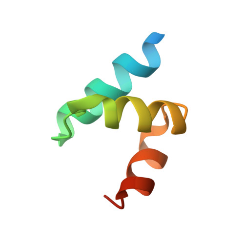
Crystal structure of the ubiquitin-associated (UBA) domain of p62 and its interaction with ubiquitin.

Isogai, S.Morimoto, D.Arita, K.Unzai, S.Tenno, T.Hasegawa, J.Sou, Y.S.Komatsu, M.Tanaka, K.Shirakawa, M.Tochio, H.

(2011) J Biol Chem 286: 31864-31874

  • DOI: https://doi.org/10.1074/jbc.M111.259630
  • Primary Citation of Related Structures:  
    3B0F

  • PubMed Abstract: 

    p62/SQSTM1/A170 is a multimodular protein that is found in ubiquitin-positive inclusions associated with neurodegenerative diseases. Recent findings indicate that p62 mediates the interaction between ubiquitinated proteins and autophagosomes, leading these proteins to be degraded via the autophagy-lysosomal pathway. This ubiquitin-mediated selective autophagy is thought to begin with recognition of the ubiquitinated proteins by the C-terminal ubiquitin-associated (UBA) domain of p62. We present here the crystal structure of the UBA domain of mouse p62 and the solution structure of its ubiquitin-bound form. The p62 UBA domain adopts a novel dimeric structure in crystals, which is distinctive from those of other UBA domains. NMR analyses reveal that in solution the domain exists in equilibrium between the dimer and monomer forms, and binding ubiquitin shifts the equilibrium toward the monomer to form a 1:1 complex between the UBA domain and ubiquitin. The dimer-to-monomer transition is associated with a structural change of the very C-terminal end of the p62 UBA domain, although the UBA fold itself is essentially maintained. Our data illustrate that dimerization and ubiquitin binding of the p62 UBA domain are incompatible with each other. These observations reveal an autoinhibitory mechanism in the p62 UBA domain and suggest that autoinhibition plays a role in the function of p62.


  • Organizational Affiliation

    Department of Molecular Engineering, Graduate School of Engineering, Kyoto University, Kyoto-Daigaku Katsura, Kyoto 615-8510, Japan.


Macromolecules
Find similar proteins by:  (by identity cutoff)  |  3D Structure
Entity ID: 1
MoleculeChains Sequence LengthOrganismDetailsImage
Sequestosome-1
A, B
53Mus musculusMutation(s): 0 
Gene Names: Sqstm1A170STAP
UniProt & NIH Common Fund Data Resources
Find proteins for Q64337 (Mus musculus)
Explore Q64337 
Go to UniProtKB:  Q64337
IMPC:  MGI:107931
Entity Groups  
Sequence Clusters30% Identity50% Identity70% Identity90% Identity95% Identity100% Identity
UniProt GroupQ64337
Sequence Annotations
Expand
  • Reference Sequence
Small Molecules
Ligands 1 Unique
IDChains Name / Formula / InChI Key2D Diagram3D Interactions
SO4
Query on SO4

Download Ideal Coordinates CCD File 
C [auth A]SULFATE ION
O4 S
QAOWNCQODCNURD-UHFFFAOYSA-L
Experimental Data & Validation

Experimental Data

  • Method: X-RAY DIFFRACTION
  • Resolution: 1.40 Å
  • R-Value Free: 0.201 
  • R-Value Work: 0.170 
  • R-Value Observed: 0.172 
  • Space Group: P 43 21 2
Unit Cell:
Length ( Å )Angle ( ˚ )
a = 72.967α = 90
b = 72.967β = 90
c = 32.56γ = 90
Software Package:
Software NamePurpose
SERGUIdata collection
SOLVEphasing
REFMACrefinement
HKL-2000data scaling

Structure Validation

View Full Validation Report


Entry History 

Deposition Data

Revision History  (Full details and data files)

  • Version 1.0: 2011-06-29
    Type: Initial release
  • Version 1.1: 2011-07-13
    Changes: Version format compliance
  • Version 1.2: 2017-10-11
    Changes: Refinement description
  • Version 1.3: 2019-11-13
    Changes: Data collection, Database references
  • Version 1.4: 2024-03-13
    Changes: Data collection, Database references, Derived calculations