3SUB

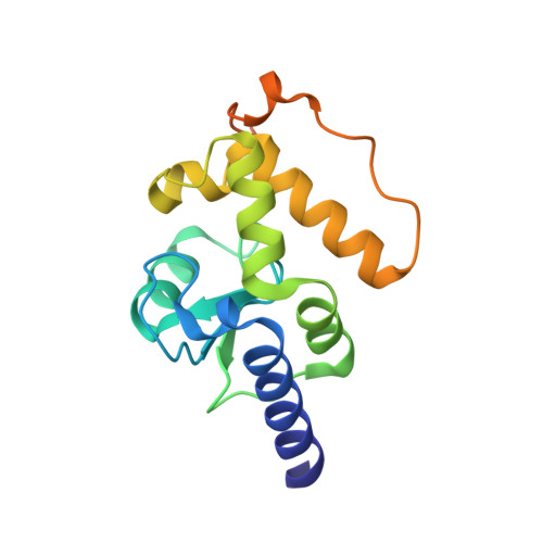
Crystal structure of the catalytic domain of Plasmodium falciparum ARF GTPase activating protein


Experimental Data Snapshot

  • Method: X-RAY DIFFRACTION
  • Resolution: 2.40 Å
  • R-Value Free: 0.227 
  • R-Value Work: 0.209 
  • R-Value Observed: 0.209 

wwPDB Validation   3D Report Full Report


This is version 1.2 of the entry. See complete history


Literature

Structure of the catalytic domain of Plasmodium falciparum ARF GTPase-activating protein (ARFGAP).

Cook, W.J.Senkovich, O.Chattopadhyay, D.

(2011) Acta Crystallogr Sect F Struct Biol Cryst Commun 67: 1339-1344

  • DOI: https://doi.org/10.1107/S1744309111032507
  • Primary Citation of Related Structures:  
    3SUB

  • PubMed Abstract: 

    The crystal structure of the catalytic domain of the ADP ribosylation factor GTPase-activating protein (ARFGAP) from Plasmodium falciparum has been determined and refined to 2.4 Å resolution. Multiwavength anomalous diffraction (MAD) data were collected utilizing the Zn(2+) ion bound at the zinc-finger domain and were used to solve the structure. The overall structure of the domain is similar to those of mammalian ARFGAPs. However, several amino-acid residues in the area where GAP interacts with ARF1 differ in P. falciparum ARFGAP. Moreover, a number of residues that form the dimer interface in the crystal structure are unique in P. falciparum ARFGAP.


  • Organizational Affiliation

    Department of Medicine, University of Alabama at Birmingham, CBSE-250, 1015 18th Street South, Birmingham, AL 35294, USA.


Macromolecules
Find similar proteins by:  (by identity cutoff)  |  3D Structure
Entity ID: 1
MoleculeChains Sequence LengthOrganismDetailsImage
ADP-ribosylation factor GTPase-activating protein
A, B
163Plasmodium falciparum 3D7Mutation(s): 0 
Gene Names: PFL2140c
UniProt
Find proteins for Q8I4Y5 (Plasmodium falciparum (isolate 3D7))
Explore Q8I4Y5 
Go to UniProtKB:  Q8I4Y5
Entity Groups  
Sequence Clusters30% Identity50% Identity70% Identity90% Identity95% Identity100% Identity
UniProt GroupQ8I4Y5
Sequence Annotations
Expand
  • Reference Sequence
Experimental Data & Validation

Experimental Data

  • Method: X-RAY DIFFRACTION
  • Resolution: 2.40 Å
  • R-Value Free: 0.227 
  • R-Value Work: 0.209 
  • R-Value Observed: 0.209 
  • Space Group: P 32 2 1
Unit Cell:
Length ( Å )Angle ( ˚ )
a = 95.9α = 90
b = 95.9β = 90
c = 92.8γ = 120
Software Package:
Software NamePurpose
HKL-2000data collection
SOLVEphasing
REFMACrefinement
HKL-2000data reduction
HKL-2000data scaling

Structure Validation

View Full Validation Report


Entry History 

Deposition Data

Revision History  (Full details and data files)

  • Version 1.0: 2011-11-09
    Type: Initial release
  • Version 1.1: 2012-03-28
    Changes: Database references
  • Version 1.2: 2024-02-28
    Changes: Data collection, Database references, Derived calculations