3V3B

Structure of the Stapled p53 Peptide Bound to Mdm2


Experimental Data Snapshot

  • Method: X-RAY DIFFRACTION
  • Resolution: 2.00 Å
  • R-Value Free: 0.216 
  • R-Value Work: 0.166 
  • R-Value Observed: 0.168 

wwPDB Validation   3D Report Full Report


This is version 2.0 of the entry. See complete history


Literature

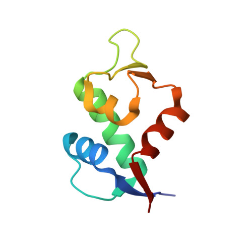
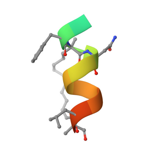
Structure of the stapled p53 peptide bound to Mdm2.

Baek, S.Kutchukian, P.S.Verdine, G.L.Huber, R.Holak, T.A.Lee, K.W.Popowicz, G.M.

(2012) J Am Chem Soc 134: 103-106

  • DOI: https://doi.org/10.1021/ja2090367
  • Primary Citation of Related Structures:  
    3V3B

  • PubMed Abstract: 

    Mdm2 is a major negative regulator of the tumor suppressor p53 protein, a protein that plays a crucial role in maintaining genome integrity. Inactivation of p53 is the most prevalent defect in human cancers. Inhibitors of the Mdm2-p53 interaction that restore the functional p53 constitute potential nongenotoxic anticancer agents with a novel mode of action. We present here a 2.0 Å resolution structure of the Mdm2 protein with a bound stapled p53 peptide. Such peptides, which are conformationally and proteolytically stabilized with all-hydrocarbon staples, are an emerging class of biologics that are capable of disrupting protein-protein interactions and thus have broad therapeutic potential. The structure represents the first crystal structure of an i, i + 7 stapled peptide bound to its target and reveals that rather than acting solely as a passive conformational brace, a staple can intimately interact with the surface of a protein and augment the binding interface.


  • Organizational Affiliation

    Max Planck Institute for Biochemistry, Am Klopferspitz 18, 82152 Martinsried, Germany.


Macromolecules
Find similar proteins by:  (by identity cutoff)  |  3D Structure
Entity ID: 1
MoleculeChains Sequence LengthOrganismDetailsImage
E3 ubiquitin-protein ligase Mdm2
A, B
88Homo sapiensMutation(s): 0 
Gene Names: MDM2
EC: 6.3.2
UniProt & NIH Common Fund Data Resources
Find proteins for Q00987 (Homo sapiens)
Explore Q00987 
Go to UniProtKB:  Q00987
PHAROS:  Q00987
GTEx:  ENSG00000135679 
Entity Groups  
Sequence Clusters30% Identity50% Identity70% Identity90% Identity95% Identity100% Identity
UniProt GroupQ00987
Sequence Annotations
Expand
  • Reference Sequence

Find similar proteins by:  Sequence   |   3D Structure  

Entity ID: 2
MoleculeChains Sequence LengthOrganismDetailsImage
SAH-p53-8 stapled-peptide
C, D
16N/AMutation(s): 0 
Entity Groups  
Sequence Clusters30% Identity50% Identity70% Identity90% Identity95% Identity100% Identity
Sequence Annotations
Expand
  • Reference Sequence
Biologically Interesting Molecules (External Reference) 1 Unique
Experimental Data & Validation

Experimental Data

  • Method: X-RAY DIFFRACTION
  • Resolution: 2.00 Å
  • R-Value Free: 0.216 
  • R-Value Work: 0.166 
  • R-Value Observed: 0.168 
  • Space Group: P 1 21 1
Unit Cell:
Length ( Å )Angle ( ˚ )
a = 45.4α = 90
b = 42.41β = 90.86
c = 50.5γ = 90
Software Package:
Software NamePurpose
MAR345dtbdata collection
MOLREPphasing
REFMACrefinement
XDSdata reduction
XSCALEdata scaling

Structure Validation

View Full Validation Report


Entry History 

Deposition Data

Revision History  (Full details and data files)

  • Version 1.0: 2012-01-18
    Type: Initial release
  • Version 1.1: 2012-03-21
    Changes: Database references
  • Version 1.2: 2012-12-12
    Changes: Other
  • Version 1.3: 2013-11-13
    Changes: Other
  • Version 2.0: 2023-11-15
    Changes: Atomic model, Data collection, Database references, Derived calculations