3VOB

Staphylococcus aureus FtsZ with PC190723


Experimental Data Snapshot

  • Method: X-RAY DIFFRACTION
  • Resolution: 2.70 Å
  • R-Value Free: 0.247 
  • R-Value Work: 0.195 
  • R-Value Observed: 0.199 

wwPDB Validation   3D Report Full Report


Ligand Structure Quality Assessment 


This is version 1.2 of the entry. See complete history


Literature

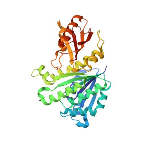
Structural reorganization of the bacterial cell-division protein FtsZ from Staphylococcus aureus

Matsui, T.Yamane, J.Mogi, N.Yamaguchi, H.Takemoto, H.Yao, M.Tanaka, I.

(2012) Acta Crystallogr D Biol Crystallogr 68: 1175-1188

  • DOI: https://doi.org/10.1107/S0907444912022640
  • Primary Citation of Related Structures:  
    3VO8, 3VO9, 3VOA, 3VOB, 3VPA

  • PubMed Abstract: 

    FtsZ is a key molecule in bacterial cell division. In the presence of GTP, it polymerizes into tubulin-like protofilaments by head-to-tail association. Protofilaments of FtsZ seem to adopt a straight or a curved conformation in relation to the bound nucleotide. However, although several bacterial and archaeal FtsZ structures have been determined, all of the structures reported previously are considered to have a curved conformation. In this study, structures of FtsZ from Staphylococcus aureus (SaFtsZ) were determined in apo, GDP-bound and inhibitor-complex forms and it was found that SaFtsZ undergoes marked conformational changes. The accumulated evidence suggests that the GDP-bound structure has the features of the straight form. The structural change between the curved and straight forms shows intriguing similarity to the eukaryotic cytoskeletal protein tubulin. Furthermore, the structure of the apo form showed an unexpectedly large conformational change in the core region. FtsZ has also been recognized as a novel target for antibacterial drugs. The structure of the complex with the inhibitor PC190723, which has potent and selective antistaphylococcal activity, indicated that the inhibitor binds at the cleft between the two subdomains.


  • Organizational Affiliation

    Faculty of Advanced Life Science, Hokkaido University, Sapporo, Hokkaido, Japan.


Macromolecules
Find similar proteins by:  (by identity cutoff)  |  3D Structure
Entity ID: 1
MoleculeChains Sequence LengthOrganismDetailsImage
Cell division protein FtsZ308Staphylococcus aureus subsp. aureus Mu50Mutation(s): 0 
Gene Names: ftsZ
UniProt
Find proteins for P0A029 (Staphylococcus aureus (strain Mu50 / ATCC 700699))
Explore P0A029 
Go to UniProtKB:  P0A029
Entity Groups  
Sequence Clusters30% Identity50% Identity70% Identity90% Identity95% Identity100% Identity
UniProt GroupP0A029
Sequence Annotations
Expand
  • Reference Sequence
Small Molecules
Ligands 3 Unique
IDChains Name / Formula / InChI Key2D Diagram3D Interactions
GDP
Query on GDP

Download Ideal Coordinates CCD File 
C [auth A]GUANOSINE-5'-DIPHOSPHATE
C10 H15 N5 O11 P2
QGWNDRXFNXRZMB-UUOKFMHZSA-N
9PC
Query on 9PC

Download Ideal Coordinates CCD File 
D [auth A]3-[(6-chloro[1,3]thiazolo[5,4-b]pyridin-2-yl)methoxy]-2,6-difluorobenzamide
C14 H8 Cl F2 N3 O2 S
INYJNSBDHOVLAH-UHFFFAOYSA-N
CA
Query on CA

Download Ideal Coordinates CCD File 
B [auth A]CALCIUM ION
Ca
BHPQYMZQTOCNFJ-UHFFFAOYSA-N
Binding Affinity Annotations 
IDSourceBinding Affinity
9PC BindingDB:  3VOB IC50: 55 (nM) from 1 assay(s)
EC50: 4000 (nM) from 1 assay(s)
Experimental Data & Validation

Experimental Data

  • Method: X-RAY DIFFRACTION
  • Resolution: 2.70 Å
  • R-Value Free: 0.247 
  • R-Value Work: 0.195 
  • R-Value Observed: 0.199 
  • Space Group: C 1 2 1
Unit Cell:
Length ( Å )Angle ( ˚ )
a = 72.095α = 90
b = 50.507β = 110.95
c = 88.288γ = 90
Software Package:
Software NamePurpose
MOLREPphasing
PHENIXrefinement
PDB_EXTRACTdata extraction
HKL-2000data collection
HKL-2000data reduction
HKL-2000data scaling

Structure Validation

View Full Validation Report


Ligand Structure Quality Assessment 


Entry History 

Deposition Data

Revision History  (Full details and data files)

  • Version 1.0: 2012-08-29
    Type: Initial release
  • Version 1.1: 2013-08-14
    Changes: Database references
  • Version 1.2: 2023-11-08
    Changes: Data collection, Database references, Derived calculations, Refinement description