4DA4

Structure of mouse DNMT1 (731-1602) bound to hemimethylated CpG DNA


Experimental Data Snapshot

  • Method: X-RAY DIFFRACTION
  • Resolution: 2.60 Å
  • R-Value Free: 0.246 
  • R-Value Work: 0.196 
  • R-Value Observed: 0.198 

wwPDB Validation   3D Report Full Report


Ligand Structure Quality Assessment 


This is version 1.1 of the entry. See complete history


Literature

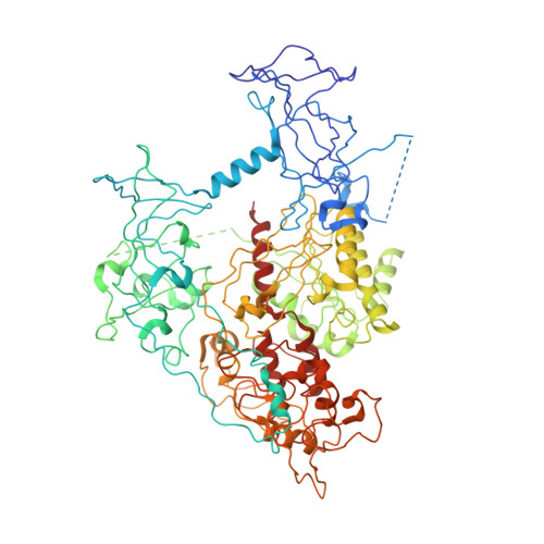
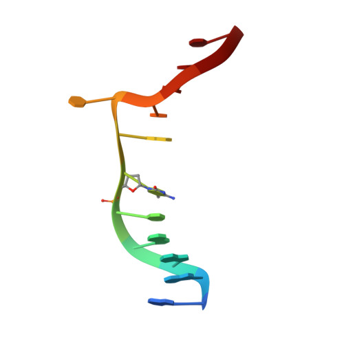
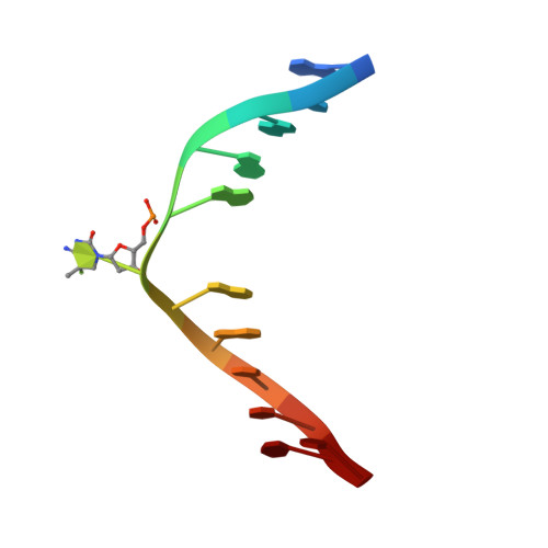
Structure-Based Mechanistic Insights into DNMT1-Mediated Maintenance DNA Methylation.

Song, J.Teplova, M.Ishibe-Murakami, S.Patel, D.J.

(2012) Science 335: 709-712

  • DOI: https://doi.org/10.1126/science.1214453
  • Primary Citation of Related Structures:  
    4DA4

  • PubMed Abstract: 

    DNMT1, the major maintenance DNA methyltransferase in animals, helps to regulate gene expression, genome imprinting, and X-chromosome inactivation. We report on the crystal structure of a productive covalent mouse DNMT1(731-1602)-DNA complex containing a central hemimethylated CpG site. The methyl group of methylcytosine is positioned within a shallow hydrophobic concave surface, whereas the cytosine on the target strand is looped out and covalently anchored within the catalytic pocket. The DNA is distorted at the hemimethylated CpG step, with side chains from catalytic and recognition loops inserting through both grooves to fill an intercalation-type cavity associated with a dual base flip-out on partner strands. Structural and biochemical data establish how a combination of active and autoinhibitory mechanisms ensures the high fidelity of DNMT1-mediated maintenance DNA methylation.


  • Organizational Affiliation

    Structural Biology Program, Memorial Sloan-Kettering Cancer Center, New York, NY 10065, USA.


Macromolecules

Find similar proteins by:  (by identity cutoff)  |  3D Structure
Entity ID: 1
MoleculeChains Sequence LengthOrganismDetailsImage
DNA (cytosine-5)-methyltransferase 1
A, B
873Mus musculusMutation(s): 0 
Gene Names: Dnmt1DnmtMet1Uim
EC: 2.1.1.37
UniProt
Find proteins for P13864 (Mus musculus)
Explore P13864 
Go to UniProtKB:  P13864
Entity Groups  
Sequence Clusters30% Identity50% Identity70% Identity90% Identity95% Identity100% Identity
UniProt GroupP13864
Sequence Annotations
Expand
  • Reference Sequence

Find similar nucleic acids by:  Sequence   |   3D Structure  

Entity ID: 2
MoleculeChains LengthOrganismImage
DNA_UPPER_STRAND
C, E
12N/A
Sequence Annotations
Expand
  • Reference Sequence

Find similar nucleic acids by:  Sequence   |   3D Structure  

Entity ID: 3
MoleculeChains LengthOrganismImage
DNA_LOWER_STRAND
D, F
12N/A
Sequence Annotations
Expand
  • Reference Sequence
Small Molecules
Binding Affinity Annotations 
IDSourceBinding Affinity
SAH BindingDB:  4DA4 IC50: min: 71, max: 4000 (nM) from 8 assay(s)
Experimental Data & Validation

Experimental Data

  • Method: X-RAY DIFFRACTION
  • Resolution: 2.60 Å
  • R-Value Free: 0.246 
  • R-Value Work: 0.196 
  • R-Value Observed: 0.198 
  • Space Group: P 1 21 1
Unit Cell:
Length ( Å )Angle ( ˚ )
a = 89.68α = 90
b = 152.042β = 94.54
c = 96.053γ = 90
Software Package:
Software NamePurpose
MAR345dtbdata collection
PHASERphasing
PHENIXrefinement
HKL-2000data reduction
HKL-2000data scaling

Structure Validation

View Full Validation Report


Ligand Structure Quality Assessment 


Entry History 

Deposition Data

Revision History  (Full details and data files)

  • Version 1.0: 2012-02-22
    Type: Initial release
  • Version 1.1: 2024-02-28
    Changes: Data collection, Database references, Derived calculations